Chromosome behaviour in mitosis: five questions to try now
Real past-paper questions, the answer key from the mark scheme, and the explanation that goes with it. No account needed to answer them.
Question 1
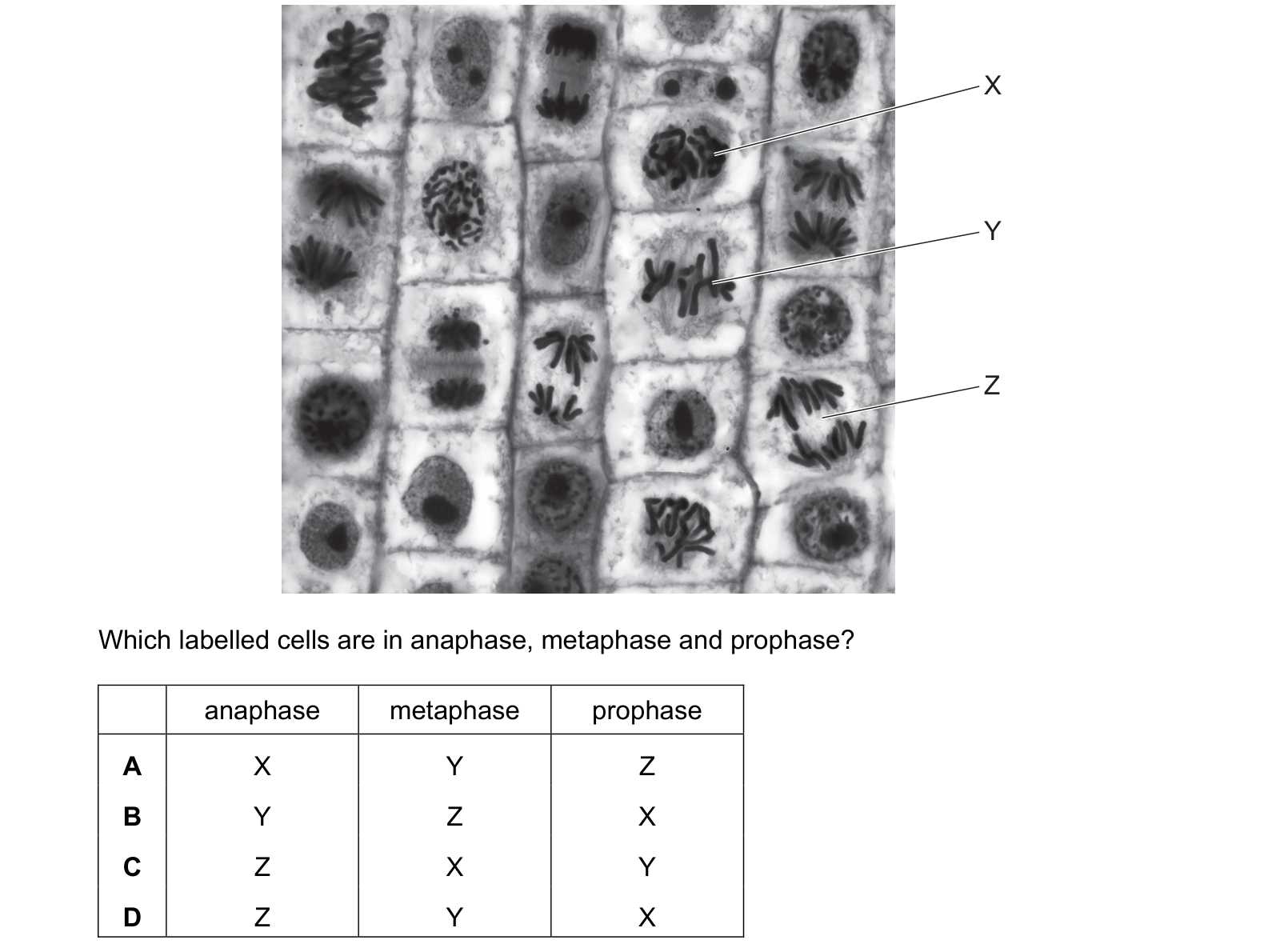
The photomicrograph shows cells in an onion root tip. Which labelled cells are in anaphase, metaphase and prophase? Each answer gives, in order: anaphase; metaphase; prophase.
Answer: D.
X is a dense tangle of thick chromosomes bunched together, still roughly filling where the nucleus was. Condensed but not organised is prophase.
Y has its chromosomes in a single line across the middle of the cell. Lined up on the equator is metaphase, and nothing else in mitosis looks like it.
Z has two separate groups of chromosomes drawn towards opposite ends, each chromosome V-shaped with the centromere leading. Two groups moving apart is anaphase.
The question asks for anaphase, metaphase, prophase in that order, so the row reads Z, Y, X, which is D.
Watch the order the columns are given in. The stages run prophase, metaphase, anaphase, and the question deliberately lists them backwards, so a candidate who identifies all three cells correctly and then writes them in the familiar order picks A and loses the mark. Read the headings before filling the row.
Question 2
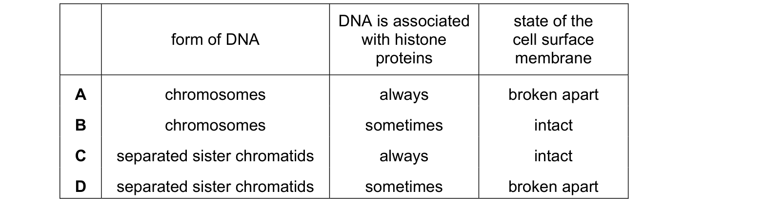
Which row is correct for the start of anaphase of mitosis? Each answer gives, in order: form of DNA; DNA is associated with histone proteins; state of the cell surface membrane.
Answer: C.
At the start of anaphase the centromeres have just divided and the sister chromatids have separated, which is what anaphase is. That settles the first column against A and B.
DNA in a eukaryotic nucleus is always associated with histone proteins, in every stage of the cycle. There is no point at which it is naked, so 'sometimes' is wrong.
The cell surface membrane is intact. Mitosis divides the nucleus; the cell does not begin to split until cytokinesis, and even then it is pinched or partitioned rather than broken.
Separated chromatids, always, intact, which is C.
Question 3
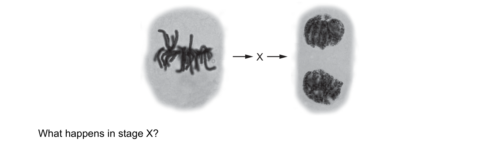
Eukaryotic organisms grow and increase in size as a result of cell division. The diagram shows some stages of mitosis with one stage missing, labelled X. What happens in stage X?
Answer: D.
That is D. The spindle fibres are microtubules, they shorten, and the centromere holding each pair of sister chromatids together divides so the two can be pulled apart.
Every wrong option is a real stage, just not this one. A is metaphase, and the first photograph shows it has already happened: the chromosomes are at the equator. B is prophase, earlier still, and it is over by the time the chromosomes are visible as tight separate bodies. C is not mitosis at all. Chromosomes lining up in pairs at the equator means homologous pairs side by side, which happens in metaphase I of meiosis. In mitosis they line up singly.
The word to watch in C is "pairs". Every mitosis question has one option describing meiosis, and that is usually how it is worded.
Question 4
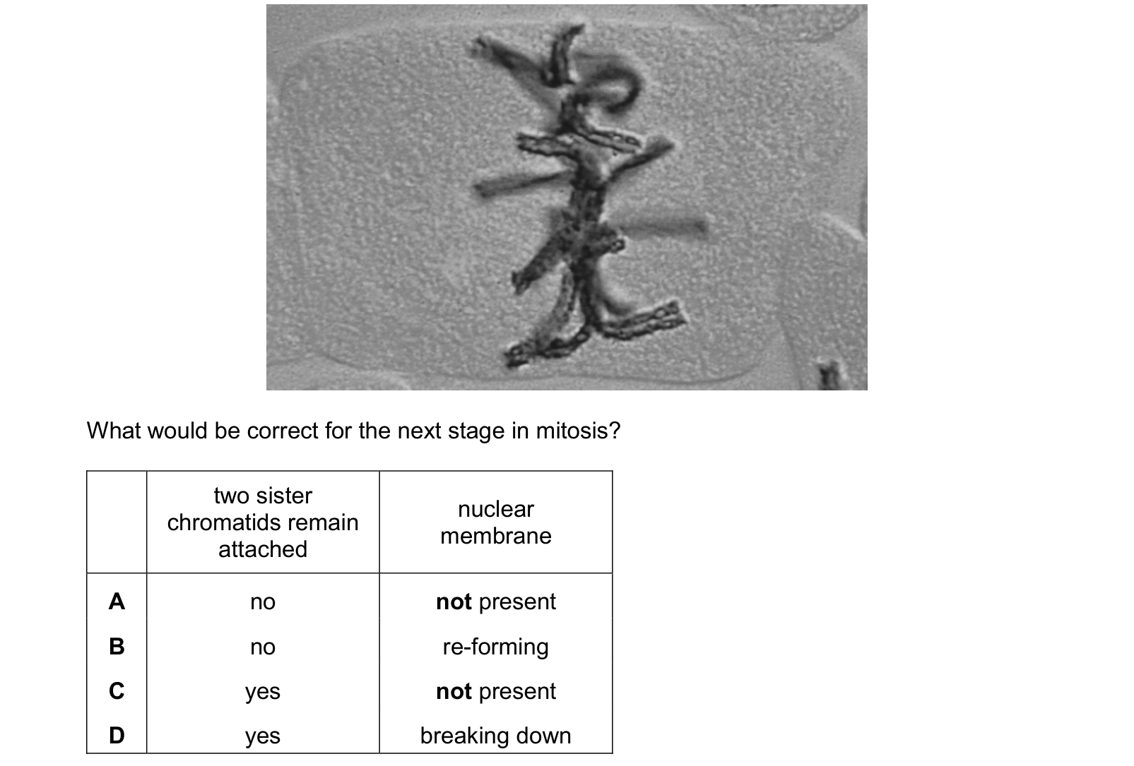
The photomicrograph shows a stage of mitosis. What would be correct for the next stage in mitosis? Each answer gives, in order: two sister chromatids remain attached; nuclear membrane.
Answer: A.
Do two sister chromatids remain attached? No. Anaphase begins precisely when the centromere divides, letting the two sister chromatids separate so one of each pair can be pulled to each pole. If they stayed attached, the daughter cells could not each receive a complete set.
Nuclear membrane: not present. It broke down during prophase, and that had to happen before the spindle could reach the chromosomes at all. It does not come back until telophase, one stage later than the one being asked about.
No, and not present, which is A.
B has the membrane re-forming, which is telophase and one stage too far. C and D both keep the chromatids attached, which would leave anaphase indistinguishable from metaphase, and D also has the membrane breaking down, which is a stage too early.
Reading the current stage off the picture is most of the work here. Everything after it follows from knowing that metaphase comes next.
Question 5
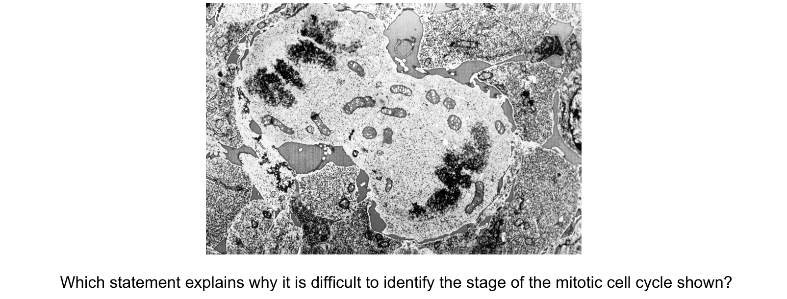
The transmission electron micrograph shows a cell in a stage of the mitotic cell cycle.
Which statement explains why it is difficult to identify the stage of the mitotic cell cycle shown?
Answer: B.
That is exactly the boundary between anaphase and telophase, which is B. Anaphase ends when the chromatids reach the poles; telophase begins as they decondense and the nuclear envelopes re-form. A single still image taken anywhere near that moment cannot say which side of it the cell is on, because the two stages are defined by a change that is gradual.
A describes something the image does not show. The chromosomes are visible, but centrioles are irrelevant here: this is a plant cell, and plant cells form a spindle without them.
C misreads the picture. There is one cell with two groups of chromosomes, not two cells. No cell wall or membrane has formed between them, which is what would be needed for two.
D is about a boundary much further on. Interphase does not begin until telophase and cytokinesis are complete, and this cell has plainly not divided yet.
The wider point is a real one about biology rather than about this photograph. The stages of mitosis are names given to a continuous process, so cells caught between two of them are common, and being unable to assign one is sometimes the correct answer.
What this practice covers
These questions are drawn from past CIE 9700 Biology papers and filtered to chromosome behaviour in mitosis. You answer, you find out immediately whether you were right, and you get the reasoning for the correct option and for each distractor. Wrong answers go to a mistakes locker so you can come back to exactly those.
Practice is free. You need an account only so your progress and your mistakes are still there next time.
What examiners see students get wrong here
These are the errors that cost marks on chromosome behaviour in mitosis, taken from our own topic notes. Read them before you practise and you will recognise the traps in the questions.
- Saying chromosomes "are made" in prophase. They exist already; they condense and become visible.
- Saying chromosomes line up in pairs at metaphase. That is meiosis. In mitosis they line up singly.
- Calling cytokinesis a stage of mitosis. Mitosis is nuclear division.
- Including interphase cells in the numerator of the mitotic index.
- Saying plant cells have no spindle because they have no centrioles. They form a spindle without them.
Revise it first
If any of the above is unfamiliar, work through the notes before practising: Chromosome behaviour in mitosis revision notes.