Chromosome behaviour in mitosis
Contents: 11 sections
Before the stages

At the start of mitosis, DNA replication has already happened in S phase. Every chromosome is therefore two identical sister chromatids joined at a centromere, and this is what makes the whole process work: the two chromatids can be separated to give two identical sets.
Chromatin is the loose, extended form the DNA is in through interphase. It only condenses into visible chromosomes when division begins, because extended DNA would tangle and break if it were dragged around the cell.
Prophase
Chromosomes condense. The chromatin coils and supercoils around histone proteins, becoming shorter and thicker until each chromosome is visible under a light microscope as two chromatids.
The nuclear envelope breaks down and the nucleolus disappears.
The spindle forms. In animal cells, the two centrioles move to opposite poles and the spindle fibres, made of microtubules, grow between them. Plant cells have no centrioles and form a spindle perfectly well, which tells you the centrioles are not essential to the process, only associated with it in animals.
Prophase is the longest stage of mitosis.
Metaphase
Chromosomes line up along the equator of the cell, in a single plane at the middle.
Spindle fibres from each pole attach to the centromere of each chromosome, so every chromosome is held by fibres from both directions. The tension of that two-way pull is what holds the chromosomes on the equator.
The cell will not proceed until every chromosome is attached. A chromosome that separated without being attached to both poles would send both chromatids to the same daughter cell.
Anaphase
The centromeres divide and the sister chromatids are pulled apart to opposite poles.
The spindle fibres shorten, dragging each chromatid centromere-first, which is why chromatids in an anaphase photomicrograph look like a V or a J with the point leading.
Once separated, each chromatid counts as a chromosome in its own right. So at anaphase in a human cell there are momentarily 92 chromosomes in one cell, 46 heading each way.
Anaphase is the shortest stage, which is worth remembering when a question gives the proportion of cells in each stage and asks which takes longest. The proportion of cells seen in a stage is proportional to how long that stage lasts.
Anaphase requires ATP, which the mitochondria clustered near the spindle supply.
Telophase
Chromosomes reach the poles and begin to uncoil back into chromatin.
A nuclear envelope re-forms around each group, the nucleolus reappears, and the spindle breaks down.
There are now two nuclei, each with the full chromosome number, genetically identical to one another and to the original.
Cytokinesis
The division of the cytoplasm. It follows telophase and is not part of mitosis, which is nuclear division only. Questions do test that distinction.
In animal cells, a ring of protein filaments just under the cell surface membrane contracts, pulling the membrane inward. A cleavage furrow deepens until the cell is pinched in two.
In plant cells, the wall makes pinching impossible. Instead, vesicles from the Golgi body line up along the equator and fuse to form a cell plate, which grows outward until it joins the existing wall. New cell wall material is laid down on both sides of it, and gaps left in it become the plasmodesmata that connect the two cells.
| Animal | Plant | |
|---|---|---|
| Centrioles | present | absent |
| Cytokinesis | cleavage furrow, inward | cell plate, outward |
| Where mitosis occurs | throughout the body | mainly at meristems |
Reading a photomicrograph
Practical questions ask you to identify the stage. The reliable tells:
- Prophase: chromosomes visible but scattered, no nuclear envelope, no alignment.
- Metaphase: chromosomes in a single line across the middle.
- Anaphase: two separate groups moving apart, chromatids V-shaped.
- Telophase: two groups at the poles, chromosomes becoming indistinct, cell beginning to divide.
The commonest error is calling metaphase anaphase. In metaphase there is one group on the equator; in anaphase there are two groups moving apart.
Mitotic index
The mitotic index is the proportion of cells in a sample that are in mitosis:
It can be given as a decimal or a percentage, and the question will say which.
Worked example
A field of view contains 60 cells. Of these, 9 are in a recognisable stage of mitosis. What is the mitotic index as a percentage?
A high mitotic index means a high proportion of cells are dividing, so the tissue is growing quickly. Root tips and shoot tips have a high index, which is why they are used for the practical. A raised index in a tissue that should be quiescent is one of the things a pathologist looks for in a tumour sample.
Cells in interphase count in the denominator, not the numerator. Interphase is not part of mitosis, and including it is the usual mistake.
The root tip practical
Root tips are used because the meristem just behind the tip is the site of active division.
The method: cut the last few millimetres of root, warm in dilute hydrochloric acid to break down the middle lamella between cells so they separate, stain with a stain that binds DNA such as acetic orcein or toluidine blue, place on a slide, and squash under a coverslip to give a single layer of cells that light can pass through.
The squash is what makes the slide readable. A block of overlapping cells shows nothing.
Common mistakes
- Saying chromosomes "are made" in prophase. They exist already; they condense and become visible.
- Saying chromosomes line up in pairs at metaphase. That is meiosis. In mitosis they line up singly.
- Calling cytokinesis a stage of mitosis. Mitosis is nuclear division.
- Including interphase cells in the numerator of the mitotic index.
- Saying plant cells have no spindle because they have no centrioles. They form a spindle without them.
Check you have it
Question 1
The photomicrograph shows a cell during mitosis.
What is happening in this cell?
1 Chromosomes are condensing.
2 Centromeres are moving to opposite poles.
3 Spindle microtubules are shortening.
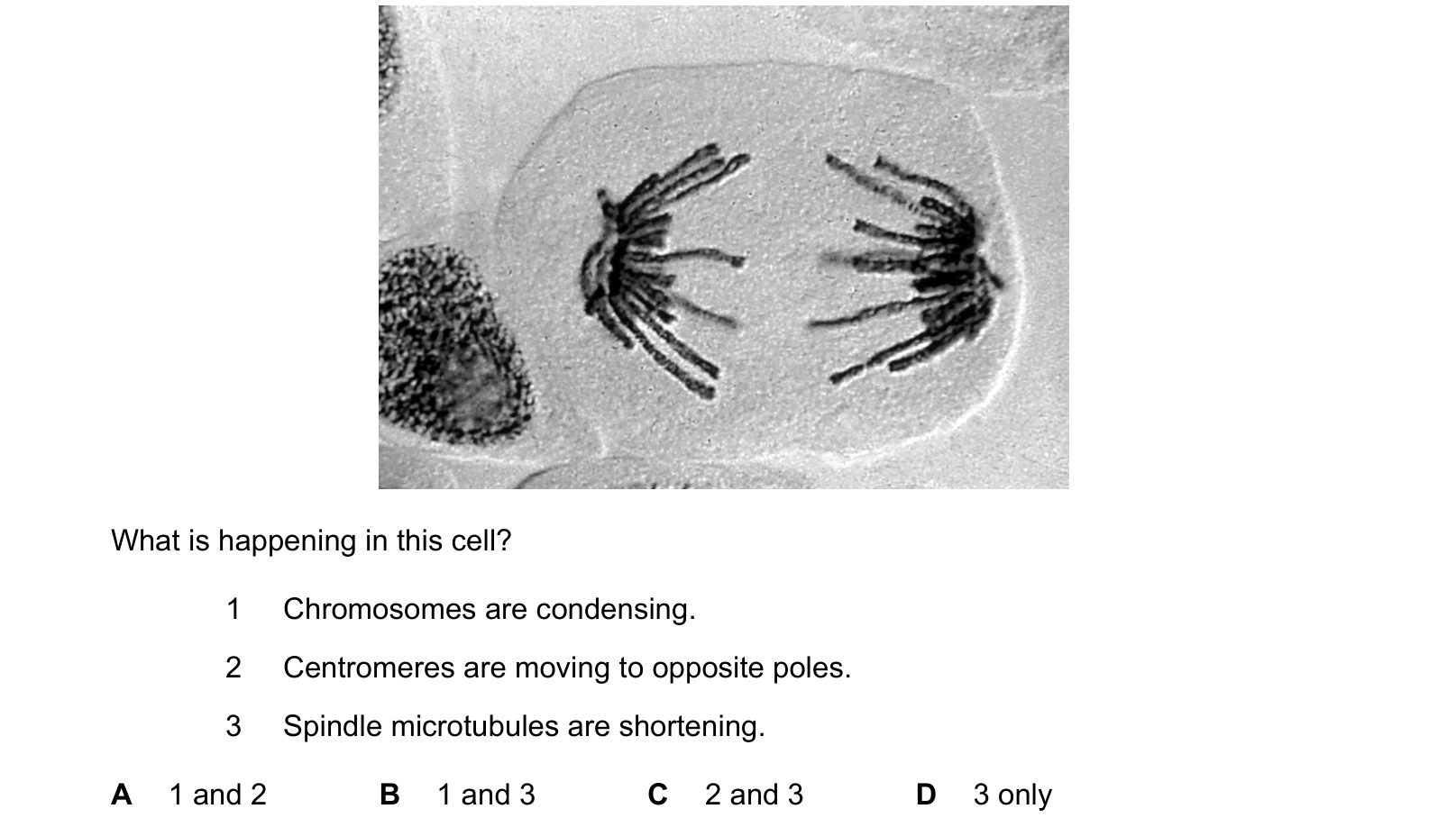
Answer: C.
2 is correct. The centromeres are moving to opposite poles, and they are what the spindle fibres are attached to.
3 is correct and it is the mechanism behind 2. The spindle fibres are microtubules, and they shorten by losing subunits at their ends, which is what reels the chromatids in. That process needs ATP, which is why cells in anaphase are rich in mitochondria near the poles.
1 is wrong. Chromosomes condense in prophase, before any of this. In the micrograph they are already fully condensed, thick enough to see individually, which is exactly why the stage is recognisable.
2 and 3, which is C.
The useful habit: name the stage from the picture first, then check each statement against that stage, rather than judging the statements one at a time in the abstract.
Question 2
The diagram shows an outline of the mitotic cell cycle. Which numbered stages of the cell cycle include a period of time when each chromosome consists of sister chromatids joined by a centromere?
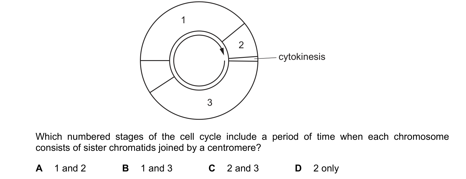
Answer: A.
Now track the chromatids. A chromosome exists as two sister chromatids joined at a centromere from the moment it is copied until the centromere divides in anaphase.
Stage 1: yes. Replication happens here, so by the end of it every chromosome is two chromatids.
Stage 2: yes. Through prophase and metaphase the chromosomes are still two chromatids each. They only become single again part way through, at anaphase, and the question asks for stages that include a period of time when the chromatids are joined, not stages where that is true throughout.
Stage 3: no. This comes after the chromatids have been separated into the two daughter cells and before the DNA is copied again, so each chromosome is a single molecule.
1 and 2, which is A.
Question 3
Which row correctly describes a stage of mitosis? Each answer gives, in order: stage of mitosis; nuclear envelope; centromeres; spindle.
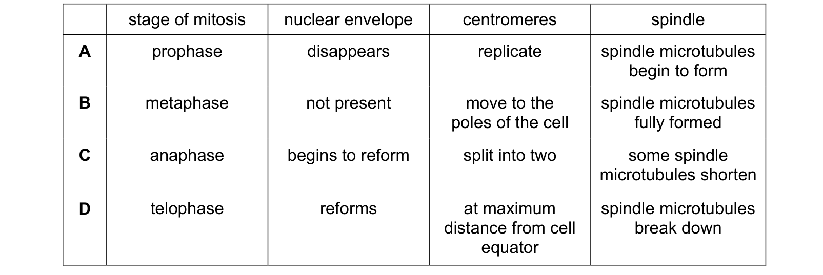
Answer: D.
D is correct throughout. In telophase the nuclear envelope re-forms around each group of chromosomes, the centromeres have been dragged to the poles so they are at maximum distance from the equator, and the spindle breaks down now that it has done its job.
A fails on one word: in prophase the centromeres do not replicate. DNA replication happened back in S phase, and prophase only makes the already-doubled chromosomes visible.
B has metaphase centromeres moving to the poles, which is anaphase. C has the nuclear envelope re-forming in anaphase, which is too early.
What the syllabus asks for on this topicSyllabus points
Syllabus points
- Describe the behaviour of chromosomes in prophase, metaphase, anaphase and telophase.
- Describe the role of the spindle and of centrioles.
- Explain how cytokinesis differs in animal and plant cells.
- Interpret photomicrographs and calculate mitotic index.
Related CIE 9700 Biology topics
Not the topic you were looking for? Describe what you are stuck on in your own words and we will take you to the notes that answer it.