The heart
Contents: 6 sections
Structure
The heart has four chambers: two thin-walled atria at the top and two thick-walled ventricles below.
The right and left sides are completely separated by the septum, which keeps oxygenated and deoxygenated blood apart. Mixing would reduce the oxygen concentration reaching the tissues.
The route through:
Deoxygenated: vena cava → right atrium → tricuspid valve → right ventricle → semilunar valve → pulmonary artery → lungs.
Oxygenated: pulmonary vein → left atrium → bicuspid valve → left ventricle → semilunar valve → aorta → body.
Why the walls differ
Atria have thin walls because they pump only into the ventricles directly below them, a very short distance.
Ventricles have thick walls because they pump blood out of the heart, and more muscle generates more force.
The left ventricle wall is two to three times thicker than the right. The right pumps to the lungs only, which are close by and whose delicate capillaries would be damaged by high pressure. The left pumps to the whole body, which needs enough pressure to reach the extremities and to still have some left after the resistance of the arterioles.
That comparison is asked constantly, and the answer that scores names both the distance and the fragility of the lung capillaries.
The valves
Atrioventricular valves sit between atrium and ventricle: the tricuspid on the right and the bicuspid (mitral) on the left. They prevent backflow from ventricle into atrium.
They are anchored by tendinous cords, tough inelastic strings attached to the ventricle wall. When ventricular pressure rises sharply the valves are forced shut, and the cords stop them being turned inside out into the atrium.
Semilunar valves sit at the exits, in the aorta and pulmonary artery, and prevent backflow from artery into ventricle.
Every valve works the same way and for the same reason: a valve opens when the pressure behind it is greater than the pressure in front, and closes when the pressure in front is greater. If you can state that sentence, every valve question in the cardiac cycle answers itself.
The coronary arteries
The heart muscle has its own blood supply, through the coronary arteries branching from the base of the aorta. The heart cannot obtain oxygen from the blood passing through its chambers, because the wall is far too thick for diffusion.
Blockage of a coronary artery starves the muscle beyond it of oxygen, and that region dies. This is a myocardial infarction.
The cardiac cycle
One complete heartbeat, lasting around 0.8 seconds at rest.
Atrial systole (about 0.1 s)
The atria contract. Pressure in the atria rises above that in the ventricles, so the atrioventricular valves are open and blood is pushed into the ventricles.
Most ventricular filling has already happened passively; atrial contraction tops it up.
Ventricular systole (about 0.3 s)
The ventricles contract. Ventricular pressure rises steeply.
- As soon as ventricular pressure exceeds atrial pressure, the atrioventricular valves close. This is the first heart sound, the "lub".
- For a moment both sets of valves are shut and the volume cannot change, so pressure rises very rapidly.
- When ventricular pressure exceeds the pressure in the aorta and pulmonary artery, the semilunar valves open and blood is ejected.
The ventricles contract from the base upward, which pushes blood towards the vessels at the top rather than trapping it at the bottom.
Diastole (about 0.4 s)
The ventricles relax and ventricular pressure falls.
- When it falls below the pressure in the arteries, the semilunar valves close. This is the second heart sound, the "dub".
- When it falls below atrial pressure, the atrioventricular valves open and blood flows passively from atria into ventricles.
The atria are filling from the veins throughout, which is why most ventricular filling happens before atrial systole.
Reading the pressure graph
The standard graph shows three lines: atrial pressure, ventricular pressure and aortic pressure, against time.
Work with the crossing points:
| Crossing | What happens |
|---|---|
| Ventricular rises above atrial | atrioventricular valve closes |
| Ventricular rises above aortic | semilunar valve opens |
| Ventricular falls below aortic | semilunar valve closes |
| Ventricular falls below atrial | atrioventricular valve opens |
Aortic pressure never falls to zero, because the elastic recoil of the aorta wall maintains it between beats. That is the small bump on the aortic line just after the semilunar valve closes.
To read heart rate from a graph, measure the time for one complete cycle and convert:
A cycle of 0.8 s gives 75 beats per minute.
Myogenic contraction and its control
The heart is myogenic: it contracts of its own accord, without a nerve impulse to start it. A heart removed from the body and supplied with oxygen and nutrients keeps beating.
The sequence:
- The sinoatrial node (SAN) in the wall of the right atrium initiates a wave of electrical excitation. It sets the rhythm, which is why it is called the pacemaker.
- The wave spreads across both atria, making them contract together.
- A layer of non-conducting tissue between the atria and ventricles stops the wave passing straight down. This is essential: it means the ventricles cannot contract at the same time as the atria.
- The wave reaches the atrioventricular node (AVN), the only route through. The AVN delays the impulse by about 0.1 seconds.
- The impulse passes down the bundle of His, through the septum to the apex at the bottom of the heart.
- It spreads up through the Purkyne tissue in the ventricle walls, and the ventricles contract from the base upward.
The two delays are the parts worth understanding rather than memorising. The AVN delay gives the atria time to finish emptying before the ventricles squeeze. The route down to the apex first means contraction starts at the bottom and pushes blood upward towards the arteries.
The rate is adjusted by nerves from the medulla, one set speeding the SAN up and the other slowing it, and by adrenaline. The heart still beats without them; they only change the rate.
Common mistakes
- Saying the left ventricle is thicker "because it pumps more blood". Both ventricles pump the same volume; the left generates more pressure.
- Saying valves open and close by muscle action. They are opened and closed by pressure differences.
- Saying the tendinous cords open the valves. They prevent them inverting.
- Saying the SAN sends a nerve impulse. It is cardiac muscle, and the wave is electrical excitation spreading through muscle.
- Forgetting why the AVN delays. Without it, the atria and ventricles would contract together and the ventricles would fill poorly.
Check you have it
Question 1
An irregular heartbeat may be the result of ineffective electrical stimulation of the atria.
Which area of the heart could be damaged, causing this irregular heartbeat?
Answer: D.
It is the pacemaker, so a fault there affects the rhythm of the whole heart, starting with the atria.
A, the atrioventricular node, sits between the atria and ventricles. Damage there delays or blocks conduction to the ventricles, leaving atrial contraction normal.
C, Purkyne tissue, carries the impulse through the ventricle walls, so again the ventricles are affected and not the atria.
B, the septum, is a wall of muscle separating left from right and does not conduct the initiating impulse.
The question says the atria, and only the SAN stimulates those first.
Question 2
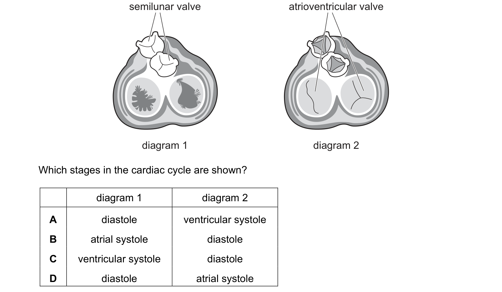
The diagrams show the valves in the heart when viewed in cross-section from above at different stages in the cardiac cycle. Which stages in the cardiac cycle are shown? Each answer gives, in order: diagram 1; diagram 2.
Answer: A.
In diagram 1 the two semilunar valves at the top are drawn shut, their cusps folded together, and the two large atrioventricular openings below are wide open. Blood is flowing from the atria into the ventricles and nothing is leaving for the arteries. The heart is filling, which is diastole.
In diagram 2 it is the other way round: the atrioventricular valves are shut, their cusps meeting across the opening, and the semilunar valves are open. The ventricles are contracting and forcing blood out into the arteries, which is ventricular systole.
Diastole then ventricular systole, which is A.
B and D both offer atrial systole, and neither diagram can be it. During atrial systole the atrioventricular valves are open and the semilunar valves are shut, which is the same valve arrangement as diagram 1, so the two are not distinguishable from a valve diagram alone. That is the trap: you cannot pick atrial systole from this evidence, so any row containing it is a guess.
The pattern behind all of it is that the two sets of valves are never open at the same time. One shuts before the other opens, which is what makes the isovolumetric phases exist.
Question 3
Heart surgery may cause a decrease in the transmission of impulses in the Purkyne tissue to the right side of the heart.
What is a possible effect of this decrease?
Answer: B.
A is about the atrioventricular node, which sits before the Purkyne tissue and is unaffected.
C is about the atria. Their contraction is triggered by the wave spreading from the sinoatrial node across the atrial muscle, which does not involve Purkyne tissue at all.
D is about the sinoatrial node, which sets the rate and is upstream of the damage.
The conduction pathway runs SAN, atria, AVN, bundle of His, Purkyne, ventricles, and damage only affects what lies downstream of it.
What the syllabus asks for on this topicSyllabus points
Syllabus points
- Describe the external and internal structure of the mammalian heart.
- Describe the cardiac cycle and explain how the valves maintain one-way flow.
- Explain how heart action is initiated and controlled by the sinoatrial node.
- Interpret pressure and volume graphs of the cardiac cycle.
Related CIE 9700 Biology topics
Not the topic you were looking for? Describe what you are stuck on in your own words and we will take you to the notes that answer it.