The gas exchange system
Contents: 6 sections
The route air takes
Trachea → bronchi → bronchioles → alveoli.
Air is drawn in through the nose and mouth, down the trachea, which divides into two bronchi, one to each lung. Each bronchus branches repeatedly into progressively narrower bronchioles, and the finest of these end in clusters of alveoli, which is where gas exchange happens.
Everything before the alveoli is conducting airway. No exchange happens there, and its job is to deliver clean, warm, moist air to the alveoli without collapsing on the way.
The tissues, and where each one is
This is the part Cambridge tests most precisely, usually as a table asking which tissue is present in which airway.
Cartilage
Present in the trachea and bronchi. Absent from bronchioles.
In the trachea it forms C-shaped rings, incomplete at the back. In the bronchi it is present as irregular blocks.
Its function is support: it holds the airway open and stops it collapsing when the pressure inside falls during inhalation. The rings are C-shaped rather than complete so that the oesophagus, immediately behind, can expand when food passes down it.
Bronchioles have no cartilage because they are held open by the surrounding lung tissue, and because cartilage would prevent them changing diameter.
Smooth muscle
Present in the trachea, bronchi and bronchioles.
It contracts to narrow the airway and relaxes to widen it, controlling the flow of air. It matters most in the bronchioles, which have no cartilage to hold them at a fixed width, so their diameter is controlled entirely by this muscle.
In asthma, the smooth muscle of the bronchioles contracts excessively, narrowing the airway and making breathing difficult.
Elastic fibres
Present throughout, including in the alveolar walls.
They stretch during inhalation as the airways and alveoli expand, and recoil during exhalation, helping to push air out. Elastic recoil is a significant part of what makes normal breathing out passive.
Emphysema destroys these fibres, so the lungs lose their recoil and exhalation becomes difficult.
Ciliated epithelium
Present in the trachea, bronchi and larger bronchioles. Absent from alveoli.
The cilia beat in a coordinated wave, sweeping mucus upward towards the throat, where it is swallowed. The mucus carries with it the dust, pollen and bacteria trapped in it. This is the mucus escalator, and it is the lung's main cleaning mechanism.
Alveoli have no cilia because a ciliated cell would be far too thick for gas exchange.
Goblet cells
Present in the trachea, bronchi and larger bronchioles. Absent from alveoli.
They secrete mucus, which traps dust and microorganisms and keeps the airway surface moist.
Goblet cells and ciliated cells work as a pair: one produces the mucus, the other moves it. Smoking paralyses the cilia, so mucus accumulates and has to be moved by coughing, which is what a smoker's cough is.
Summary table
| Trachea | Bronchus | Bronchiole | Alveolus | |
|---|---|---|---|---|
| Cartilage | yes, C-rings | yes, blocks | no | no |
| Smooth muscle | yes | yes | yes | no |
| Elastic fibres | yes | yes | yes | yes |
| Ciliated epithelium | yes | yes | larger only | no |
| Goblet cells | yes | yes | larger only | no |
Two rows carry most of the marks: cartilage stops at the bronchi, and cilia and goblet cells stop before the alveoli.
The alveoli
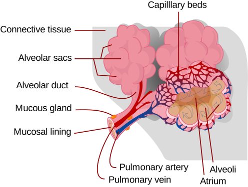
Each lung contains roughly 300 million alveoli. Every feature is about making diffusion fast, and Fick's law is the frame to hang them on: rate depends on surface area and concentration difference, divided by distance.
A very large surface area. The alveoli together give a surface of around 70 m², which is more area for diffusion.
A very short diffusion distance. The alveolar wall is a single layer of squamous epithelium, extremely flattened. The capillary wall is a single layer of endothelium. Together they are less than 1 µm thick, so oxygen crosses two thin cells and no more.
A steep concentration gradient, maintained by two things at once:
- ventilation constantly replaces the air in the alveoli, so the oxygen concentration inside stays high and the carbon dioxide concentration stays low
- a good blood supply constantly removes oxygenated blood and brings deoxygenated blood, so the gradient across the wall is never allowed to disappear
A question asking how the gradient is maintained is asking for both of those, not one.
A moist lining. The alveolar surface is coated with a thin film of fluid, and gases must dissolve before they can diffuse across a membrane.
Capillaries in close contact. The capillary network wraps the alveolus so closely that red cells are pressed against the wall, shortening the diffusion path further, and they travel slowly, allowing time for exchange.
Surfactant. A film of phospholipid on the alveolar surface reduces surface tension. Without it, the water lining would pull the alveolus closed as it emptied. Premature babies are sometimes born before they can make enough surfactant, which is why their lungs are prone to collapse.
Elastic fibres in the wall let the alveolus expand on inhalation and recoil on exhalation.
Which way each gas goes
In the alveolus, the partial pressure of oxygen is high and in the blood arriving it is low, so oxygen diffuses into the blood.
The partial pressure of carbon dioxide is high in the arriving blood and low in the alveolus, so carbon dioxide diffuses out of the blood.
Both are simple diffusion down a gradient. No energy is used, and there is no active transport in the lung.
Common mistakes
- Saying bronchioles contain cartilage. They do not, and this is the single most tested fact in the topic.
- Saying alveoli have goblet cells or cilia. They have neither, because both would be too thick.
- Saying the alveolar wall is "thin" without saying it is one cell thick.
- Saying a large surface area alone explains fast exchange. The short distance and the maintained gradient matter just as much.
- Saying ventilation maintains the gradient, and stopping there. The blood supply is the other half.
Check you have it
Question 1
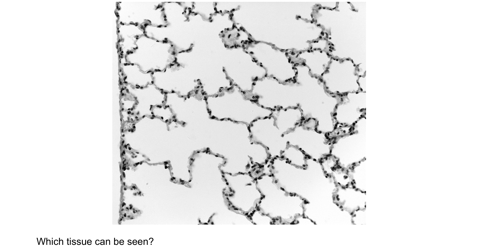
The photomicrograph shows a section through part of the gas exchange system.
Which tissue can be seen?
Answer: D.
Squamous means flattened, and here that is the whole point rather than an incidental fact. A gas has to diffuse across the alveolar wall and then across the capillary wall, and the rate of diffusion depends on the distance. Each of those layers is one flattened cell thick, so the barrier is well under a micrometre.
A, cartilage, would appear as a thick pale block with cells sitting in small spaces, and it belongs in the trachea and bronchi. There is none here.
B, ciliated epithelium, is tall columnar cells packed side by side with a dense mat of cilia along the top. Nothing in this section is tall, and there are no cilia. Cilia belong on the airways that move mucus, and putting them in an alveolus would obstruct the very barrier that has to stay thin.
C, smooth muscle, would be a band of long spindle-shaped cells running in one direction, which is not what the lacy network shows.
Question 2
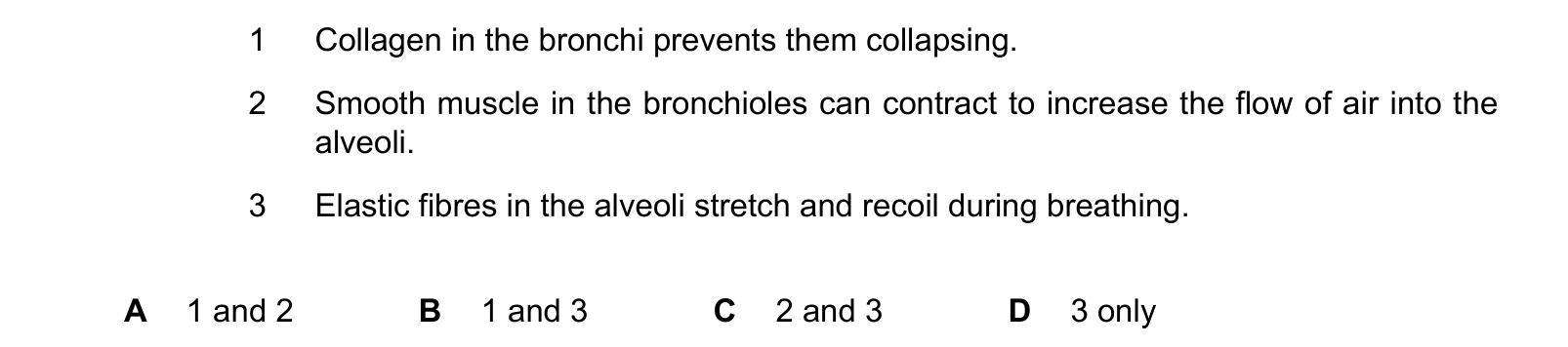
Which statements about the function of tissues found in the human gas exchange system are correct?
Answer: D.
1 names the wrong tissue. What holds the bronchi open is cartilage, in irregular plates in the bronchus and C-shaped rings in the trachea. Collagen is present in the airway walls, but the tissue that stops them collapsing as the pressure inside drops during inhalation is cartilage.
2 has the effect backwards. Smooth muscle contracting makes a bronchiole narrower, which reduces the flow of air. That is exactly what happens in an asthma attack. Air flow is increased when the muscle relaxes and the lumen widens.
3 is correct. The alveolar walls contain elastic fibres. They stretch as the alveoli fill during inhalation and recoil as the lungs empty, and that recoil is what drives passive exhalation. Losing it is what makes emphysema so disabling.
3 only, which is D.
All three statements pair a real tissue with a real place, which is what makes this harder than it looks. Check the tissue and then check the direction of the effect, separately.
Question 3
Which statements about all bronchioles are correct?
1 They have goblet cells.
2 They have epithelial cells.
3 They have muscle tissue.
Answer: D.
Every bronchiole is lined with epithelial cells, and every bronchiole has smooth muscle, which is how the airway narrows and widens.
Statement 1 fails on the word all. Goblet cells are present in the larger bronchioles and absent from the smallest ones, because mucus produced in the finest airways could not be cleared and would block them.
This is the same fact as the cilia question from the other direction: goblet cells and ciliated cells both thin out as the airways get finer, and disappear entirely before the alveoli.
What the syllabus asks for on this topicSyllabus points
Syllabus points
- Describe the structure of the human gas exchange system.
- Recognise and describe the distribution of cartilage, ciliated epithelium, goblet cells, smooth muscle and elastic fibres.
- Relate the structure of the alveoli to gas exchange.
- Describe the functions of the airway tissues.
Related CIE 9700 Biology topics
Not the topic you were looking for? Describe what you are stuck on in your own words and we will take you to the notes that answer it.