Cells as the basic units of living organisms
Contents: 9 sections
Why the electron microscope changed the answer
A light microscope shows the nucleus, the cell wall, chloroplasts and a large vacuole, and essentially nothing else. Everything else in this topic, the internal membranes, ribosomes, the structure of a mitochondrion, was invisible until the electron microscope resolved to about 0.5 nm.
So when a question specifies "as seen with an electron microscope", it is asking for the detailed answer, and organelles such as ribosomes and rough endoplasmic reticulum are expected.
The organelles
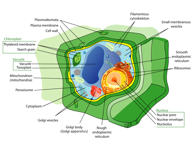
| Organelle | Structure | Function |
|---|---|---|
| Nucleus | Surrounded by a double membrane, the nuclear envelope, pierced by nuclear pores. Contains chromatin and a nucleolus. | Holds the DNA. Controls the cell by controlling protein synthesis. |
| Nucleolus | Dense region inside the nucleus, no membrane. | Makes ribosomes. |
| Rough ER | Flattened membrane sacs with ribosomes on the surface. | Transports the proteins made by its ribosomes. |
| Smooth ER | Flattened sacs, no ribosomes. | Makes lipids and steroids. |
| Golgi body | Stack of flattened sacs, with vesicles pinching off. | Modifies and packages proteins, then sends them out in vesicles. Makes lysosomes. |
| Mitochondrion | Double membrane; the inner one folded into cristae, enclosing the matrix. | Aerobic respiration, producing ATP. |
| Ribosome | No membrane. Two subunits. 80S in eukaryotes, 70S in prokaryotes. | Protein synthesis. |
| Lysosome | Single membrane sac of hydrolytic enzymes. | Digests worn-out organelles and material taken in by the cell. |
| Centriole | Two bundles of microtubules at right angles. | Makes the spindle in cell division. Absent from higher plant cells. |
| Chloroplast | Double membrane; internal thylakoids stacked into grana, surrounded by stroma. | Photosynthesis. |
| Cell wall | Cellulose in plants. Outside the membrane. | Support and shape. Stops the cell bursting. |
| Vacuole | Large and permanent in plant cells, surrounded by the tonoplast. | Stores cell sap. Keeps the cell turgid. |
The structures that carry marks
Mitochondria and chloroplasts share a pattern worth noticing. Both have a double membrane, and both have an inner membrane folded or stacked to give a very large surface area, cristae in one and thylakoids in the other. In both cases the reason is the same: the reactions are catalysed by enzymes and carriers embedded in that membrane, so more membrane means more reaction can happen at once.
Both also contain 70S ribosomes and their own circular DNA, which is the same combination found in prokaryotes. That observation is the basis of the endosymbiotic theory, and it is why "70S ribosomes" can be an acceptable answer to a question asking what a prokaryotic cell and a eukaryotic cell have in common.
Animal against plant cells
Present in plant cells and absent from animal cells:
- Cell wall of cellulose
- Chloroplasts (not in every cell: root cells have none)
- Large permanent vacuole with a tonoplast
- Starch grains as the storage carbohydrate
Present in animal cells and absent from higher plant cells:
- Centrioles
- Glycogen granules as the storage carbohydrate
- Lysosomes are common and prominent; plant cells have them but they are far less conspicuous
Both have a cell surface membrane, nucleus, mitochondria, ribosomes, rough and smooth ER, and a Golgi body. A question asking what plant and animal cells have in common is asking for these.
Prokaryotic against eukaryotic cells
| Feature | Prokaryotic | Eukaryotic |
|---|---|---|
| Nucleus | No true nucleus, no nuclear envelope | Nucleus with a double membrane |
| DNA | Circular, naked, in the cytoplasm | Linear, associated with histone proteins |
| Ribosomes | 70S | 80S in the cytoplasm, 70S in mitochondria and chloroplasts |
| Membrane-bound organelles | Absent | Present |
| Cell wall | Peptidoglycan | Cellulose in plants, chitin in fungi, absent in animals |
| Plasmids | Often present | Absent |
| Size | About 1 to 5 µm | About 20 to 100 µm |
The defining difference is the absence of membrane-bound organelles, not just the absence of a nucleus. A prokaryote has no mitochondria, no ER and no Golgi, and respires using its cell surface membrane instead. Saying only "no nucleus" answers half the question.
Note the ribosome row carefully. It is the reason 70S ribosomes appear in both cell types, and it is a favourite multiple-choice question.
Viruses
Viruses are not cells and are not counted as living organisms, because they have no cytoplasm, no organelles and no metabolism of their own. They can only replicate inside a host cell.
All viruses have:
- Nucleic acid, either DNA or RNA, never both.
- A capsid, a protein coat built from repeating subunits.
Some also have an envelope of membrane taken from the host cell as the virus leaves it. HIV and influenza have one; the tobacco mosaic virus does not.
Viruses are measured in nanometres, typically 20 to 300 nm, which places them below the resolution of a light microscope. They were not seen until the electron microscope existed, although their existence had been inferred long before from the fact that infectious material passed through filters fine enough to stop every known bacterium.
Common exam mistakes
- Answering "no nucleus" alone for the prokaryote difference, and omitting the absence of membrane-bound organelles.
- Saying prokaryotes have no ribosomes. They have ribosomes, and those ribosomes are 70S.
- Giving centrioles to plant cells, or chloroplasts to every plant cell including root cells.
- Calling the nuclear envelope a single membrane. It is double, and it is pierced by pores.
- Saying a virus is a small cell, or that a virus contains both DNA and RNA.
- Describing the function of the Golgi as "storage". It modifies, packages and exports.
Exam technique
- When asked to describe an organelle, give structure and function together. "Mitochondrion: double membrane with the inner folded into cristae, site of aerobic respiration" answers both halves of most questions.
- Where a structure has a large surface area, say what the area is for. Surface area alone is a description; linking it to the enzymes or carriers embedded there is the explanation.
- In comparison questions, write the two sides of each point. "Prokaryotes have 70S ribosomes whereas eukaryotes have 80S" scores where "prokaryotes have 70S ribosomes" may not.
- Use the sizes. A bacterium at 1 to 5 µm against a eukaryotic cell at 20 to 100 µm is a concrete comparison.
Quick revision
- Double membranes: nucleus, mitochondrion, chloroplast.
- Cristae and thylakoids both exist to increase membrane surface area for embedded enzymes and carriers.
- Mitochondria and chloroplasts contain 70S ribosomes and circular DNA, as prokaryotes do.
- Prokaryote: no nucleus and no membrane-bound organelles; circular naked DNA; 70S ribosomes; peptidoglycan wall.
- Plant only: cellulose wall, chloroplasts, large permanent vacuole. Animal only: centrioles.
- Viruses: nucleic acid plus a capsid, sometimes an envelope; 20 to 300 nm; not living.
Check you have it
Question 1
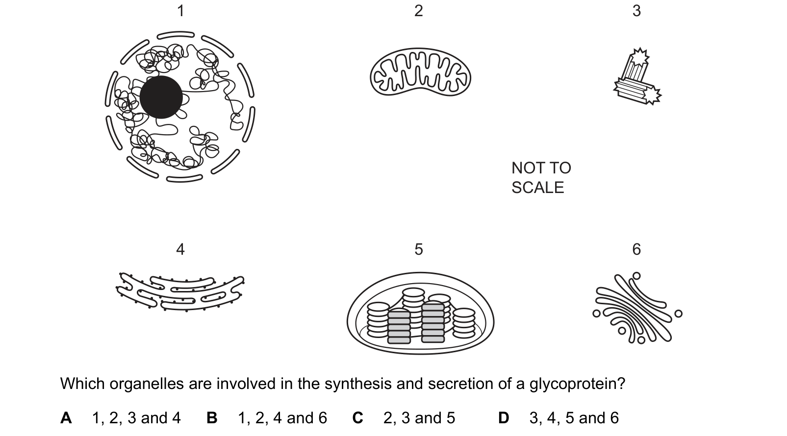
Six organelles found in eukaryotic cells are shown.
1 2 3 Which organelles are involved in the synthesis and secretion of a glycoprotein?
Answer: B.
1 is the nucleus, recognisable by its envelope and dark nucleolus. The gene is transcribed here into mRNA.
4 is the rough endoplasmic reticulum, the flattened sacs studded with ribosomes. The protein is translated by those ribosomes and fed straight into the RER as it is made.
6 is the Golgi body, curved stacks with vesicles budding off. This is the step that makes it a glycoprotein: the Golgi adds the carbohydrate chains, then packages the finished molecule into a vesicle for secretion.
2 is the mitochondrion, and it belongs on the list because every stage of this costs ATP: transcription, translation, vesicle transport and exocytosis.
1, 2, 4 and 6, which is B.
3 is a centriole, a barrel of microtubules used to organise the spindle in cell division, with no part in secretion. 5 is a chloroplast, identifiable by its stacks of grana, and it is the odd one out twice over: it does photosynthesis, and it would not be in the same cell as a secretory pathway of this kind anyway.
Question 2
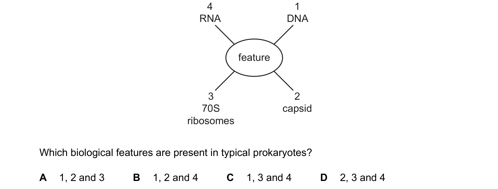
The diagram shows four biological features.
4 1 70S capsid ribosomes
Which biological features are present in typical prokaryotes?
Answer: C.
2, the capsid, is a virus feature. It is the protein coat that encloses a virus's nucleic acid, and no cell has one. That removes A, B and D in a single step, and the question is done.
Worth confirming the other three anyway.
1 DNA: yes. A prokaryote has a single circular DNA molecule lying free in the cytoplasm, with no nuclear envelope around it, and often plasmids as well.
3 70S ribosomes: yes, and the number is the point. Prokaryotic ribosomes are 70S and eukaryotic cytoplasmic ribosomes are 80S. The 70S ones inside mitochondria and chloroplasts are one of the strongest pieces of evidence that those organelles began as free-living bacteria.
4 RNA: yes. Every cell that makes protein needs mRNA, tRNA and the rRNA in its own ribosomes.
1, 3 and 4, which is C.
Question 3
Which statements about viruses are correct?
1 They contain DNA or RNA.
2 They use host cells to synthesise virus proteins.
3 They can have a protective coat of peptidoglycan.
Answer: B.
A virus contains DNA or RNA, never both, which is why the genetic material is described that way: statement 1. It has no ribosomes and no metabolism, so it uses the host cell's machinery to make its proteins: statement 2.
Statement 3 names the wrong material. A virus's coat is the capsid, made of protein. Peptidoglycan is what a bacterial cell wall is made of, and it is the target of penicillin.
That distinction matters beyond this question: it is why antibiotics do nothing against a virus. There is no peptidoglycan wall to attack and no bacterial ribosome to block.
What the syllabus asks for on this topicSyllabus points
Syllabus points
- Describe the structure of a typical animal cell and a typical plant cell as seen with an electron microscope.
- State the functions of the organelles and structures within them.
- Outline the key differences between prokaryotic and eukaryotic cells.
- Outline the structure of viruses.
Related CIE 9700 Biology topics
Not the topic you were looking for? Describe what you are stuck on in your own words and we will take you to the notes that answer it.