The circulatory system
Contents: 6 sections
Why a transport system is needed
A single-celled organism exchanges everything it needs across its surface, and diffusion is fast enough because the distances are tiny.
As an organism gets larger the surface area to volume ratio falls, so there is less surface per unit of tissue to supply. At the same time the diffusion distance to the centre increases. Diffusion over a long distance is very slow, so it cannot keep pace with the metabolic demands of an active animal.
The answer is a mass transport system: a fluid moved in bulk by a pump, delivering substances close to every cell so that the final step by diffusion is short.
A closed, double circulation
Closed means the blood stays inside vessels at all times and does not bathe the tissues directly. The advantages are that pressure can be kept high, and flow can be directed to where it is needed.
Double means the blood passes through the heart twice for each complete circuit:
- the pulmonary circulation takes deoxygenated blood from the right side of the heart to the lungs and returns oxygenated blood to the left side
- the systemic circulation takes oxygenated blood from the left side to the body and returns deoxygenated blood to the right side
The advantage of a double circulation is that blood returning from the lungs at low pressure is repressurised before being sent to the body. A single circulation, as in a fish, loses most of its pressure crossing the gill capillaries, so blood reaches the body slowly. A double circulation supports the high metabolic rate of a mammal.
Pulmonary vessels are the exception to the usual rule: the pulmonary artery carries deoxygenated blood and the pulmonary vein carries oxygenated blood. Arteries are defined by carrying blood away from the heart, not by what is in them.
The three vessel types
All three have a lumen and a wall, but the proportions differ completely, and every difference follows from the pressure the vessel handles.
Arteries
Carry blood away from the heart at high pressure, in pulses.
- Thick wall overall, to withstand the pressure without bursting.
- A thick layer of elastic fibres. These stretch as the surge of blood from the heart arrives, then recoil as it passes. That recoil pushes the blood onward and smooths the flow, so blood arrives at the capillaries as a steady stream rather than in bursts. It is also what produces a pulse you can feel.
- A thick layer of smooth muscle, which contracts to narrow the lumen and so redirects blood between organs.
- A narrow lumen relative to the wall, which helps maintain the pressure.
- Endothelium lining the lumen, one cell thick and smooth, reducing friction.
Arterioles are smaller, with proportionally more smooth muscle and less elastic tissue. They are where the fine control of blood distribution happens: contracting the muscle narrows them and reduces flow to that region.
Veins
Carry blood towards the heart at low pressure.
- Thin wall, because there is little pressure to resist.
- Little elastic tissue and little muscle, since there is no surge to smooth out and no need for the vein to recoil.
- A wide lumen, which reduces resistance to the slow-moving blood.
- Semilunar valves along their length, which prevent backflow.
Blood in veins is moved mainly by the contraction of the skeletal muscles surrounding them, which squeeze the vein, and by pressure changes during breathing. The valves ensure that squeezing a vein moves blood only towards the heart. Without them, muscle contraction would push blood equally in both directions and achieve nothing.
Capillaries
The site of exchange with the tissues.
- Wall one cell thick, just endothelium, giving the shortest possible diffusion distance.
- A very narrow lumen, roughly the diameter of a red blood cell, so red cells are squeezed against the wall and the diffusion path is shorter still.
- Gaps between the endothelial cells, allowing plasma and white blood cells through.
- Enormous total surface area, because there are so many, and no cell is far from one.
- Slow flow, because the total cross-sectional area of all the capillaries is much greater than that of the artery feeding them. Slow flow allows more time for exchange.
That last point is counterintuitive and gets asked: an individual capillary is tiny, but there are so many in parallel that the combined area is large, so the blood slows down.
Compared
| Artery | Vein | Capillary | |
|---|---|---|---|
| Direction | away from heart | towards heart | between the two |
| Pressure | high, pulsing | low, steady | falling |
| Wall | thick, elastic and muscular | thin | one cell |
| Lumen | narrow | wide | very narrow |
| Valves | no | yes | no |
Tissue fluid
Tissue fluid is the fluid that surrounds the body's cells, and it is where the actual exchange between blood and cell happens.
At the arteriole end of a capillary the blood is still at relatively high hydrostatic pressure. That pressure forces water and small dissolved molecules such as glucose, amino acids, oxygen and mineral ions out through the gaps in the capillary wall. This is ultrafiltration.
Plasma proteins are too large to leave, so they stay in the capillary. This matters because they lower the water potential of the blood plasma, and that effect stays behind when the water goes.
Along the capillary, hydrostatic pressure falls, because fluid has left and because of resistance.
At the venule end, the hydrostatic pressure is now low, but the plasma proteins are still there and are now more concentrated. The water potential of the blood is therefore lower than that of the tissue fluid, so water moves back into the capillary by osmosis, carrying dissolved waste such as carbon dioxide with it.
Not all of it returns. Around ten per cent remains in the tissues and is drained away by the lymphatic system, which returns it to the blood near the heart.
When it goes wrong
If plasma protein levels fall, through malnutrition or liver disease, the water potential of the blood is no longer low enough to draw water back at the venule end. Fluid accumulates in the tissues, which is oedema. The same happens if the lymphatic system is blocked.
Understanding oedema as a failure of one of the two pressures is a good test of whether the mechanism is really understood.
Common mistakes
- Saying arteries carry oxygenated blood. They carry blood away from the heart; the pulmonary artery is deoxygenated.
- Saying veins have valves "to keep the pressure high". Valves prevent backflow.
- Saying elastic tissue in an artery lets it stretch, and stopping there. The recoil is what maintains flow between heartbeats.
- Saying tissue fluid returns to the capillary because of hydrostatic pressure. It returns by osmosis, because plasma proteins remain behind.
- Saying blood flows slowly in capillaries because they are narrow. It is the large total cross-sectional area of all of them together.
Check you have it
Question 1
Which row is correct for an artery? Each answer gives, in order: inner layer; middle layer; outer layer.
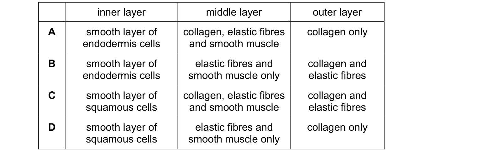
Answer: C.
The inner layer is endothelium, a smooth layer of flattened, or squamous, cells. That eliminates A and B, which call it endodermis, a term from embryology that has nothing to do with blood vessels.
The middle layer contains collagen, elastic fibres and smooth muscle together. Elastic fibres give the recoil, smooth muscle changes the diameter, and collagen limits how far the vessel can stretch.
The outer layer is collagen and elastic fibres, giving strength and preventing over-expansion.
So C. D omits the collagen from both the middle and outer layers, which would leave the artery able to stretch without limit.
Question 2
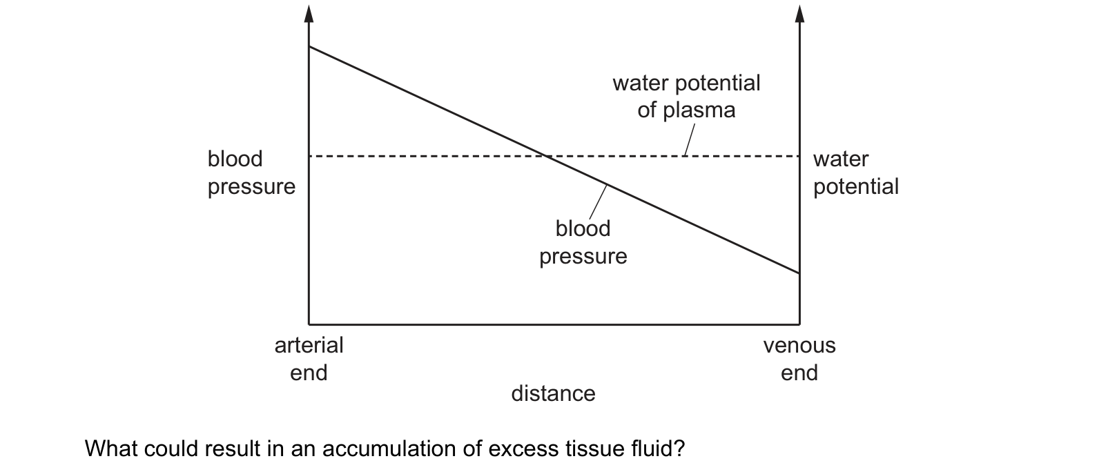
The graph shows the changes in blood pressure and water potential across a blood capillary from the arterial end of the capillary to the venous end of the capillary. end end distance
What could result in an accumulation of excess tissue fluid?
Answer: B.
B does that. Raising the blood pressure lifts the whole sloping line, so it crosses later and spends more of the capillary above the water potential line. More fluid is forced out along a longer stretch and less is drawn back in the short stretch that remains.
D is the same reasoning in reverse and would reduce tissue fluid, not increase it.
A goes the wrong way too. More large protein in the plasma makes the plasma's water potential more negative, which lowers the dashed line, moves the crossing earlier and pulls more fluid back. It is the loss of plasma protein that causes swelling, which is why severe protein deficiency and liver disease both produce oedema.
C also reduces tissue fluid. Fewer small proteins outside makes the tissue fluid's water potential less negative, so the gradient drawing water back into the capillary is steeper.
Raised blood pressure is the everyday cause: it is why ankles swell after standing all day, when gravity has raised the pressure in the capillaries of the legs.
Question 3
The diagram shows a transverse section through an artery. Which tissues are present in layer X?

Answer: D.
Each one is there for a reason worth being able to state.
Elastic fibres stretch when the ventricle ejects blood and recoil in diastole, which stores energy from each beat and keeps the blood moving between them.
Smooth muscle contracts to narrow the lumen, which is how the body directs blood to some organs and away from others.
Collagen is the one people leave out, and it is the one stopping the vessel bursting. It has enormous tensile strength and almost no stretch, so it sets a limit on how far the elastic fibres can take the wall.
A, B and C each drop one of the three. The outer layer, the tunica externa, is mostly collagen, and the innermost lining is the single layer of endothelium, neither of which is what X marks.
What the syllabus asks for on this topicSyllabus points
Syllabus points
- Explain the need for a transport system in large multicellular organisms.
- Describe the mammalian circulatory system as closed and double.
- Relate the structure of arteries, veins and capillaries to their functions.
- Describe the formation of tissue fluid and its return to the blood.
Related CIE 9700 Biology topics
Not the topic you were looking for? Describe what you are stuck on in your own words and we will take you to the notes that answer it.