The circulatory system: five questions to try now
Real past-paper questions, the answer key from the mark scheme, and the explanation that goes with it. No account needed to answer them.
Question 1
Which reactions will be taking place in blood in a capillary that is next to an alveolus?
1 Hb + 4O2 → HbO8 key
2 H2O + CO2 → H2CO3 Hb = haemoglobin
3 H2CO3 → H+ + HCO3 –

Answer: B.
So haemoglobin combining with oxygen, statement 1, is exactly what happens: Hb + 4O₂ → HbO₈. That is B.
Statement 2, carbon dioxide and water forming carbonic acid, is the loading reaction of respiring tissues. In the lungs it runs the other way, breaking carbonic acid down so the carbon dioxide can leave.
Statement 3, carbonic acid dissociating into hydrogen and hydrogencarbonate ions, is the same story: in the lungs those ions recombine instead.
The reliable method for this whole topic is to establish where the blood is first. Every reaction is reversible and the location decides the direction.
Question 2
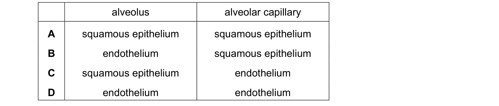
Which layers of cells does an oxygen molecule diffuse through when moving from an alveolus into an alveolar capillary? Each answer gives, in order: alveolus; alveolar capillary.
Answer: C.
Both are a single layer of extremely flattened cells, which is why the total diffusion distance is under a micrometre. What differs is the name, and the naming rule is worth knowing: epithelium lines surfaces exposed to the outside, including the airways and the gut, while endothelium lines the inside of blood and lymph vessels.
An alveolus is open to the atmosphere, so its lining is epithelium. A capillary carries blood, so its lining is endothelium.
Options A and D use the same word for both, and B has them the wrong way round.
Question 3
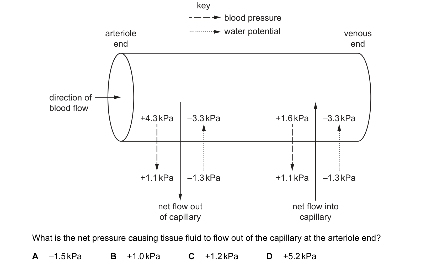
The diagram shows how tissue fluid is formed.
key direction of blood flow of capillary capillary
What is the net pressure causing tissue fluid to flow out of the capillary at the arteriole end?
Answer: C.
Pushing out at the arteriole end: the blood pressure inside, +4.3 kPa. Pushing in: the pressure of the tissue fluid outside, +1.1 kPa. Net hydrostatic effect: 4.3 − 1.1 = +3.2 kPa outwards.
Water potential is −3.3 kPa inside and −1.3 kPa outside. Water moves from higher to lower water potential, and −1.3 is higher than −3.3, so osmosis pulls water in with a strength of 3.3 − 1.3 = 2.0 kPa.
Net outward pressure = 3.2 − 2.0 = +1.2 kPa, which is C.
A is negative, and the diagram itself rules it out: the arrow at the arteriole end is labelled net flow out of the capillary, so the answer has to be positive. Check that before doing any arithmetic.
B, +1.0 kPa, uses only the two figures inside the capillary, 4.3 − 3.3, and forgets the tissue fluid entirely. D, +5.2 kPa, adds all four with the signs mixed up.
The useful habit is to write the four values in two columns, out and in, before subtracting. The same four numbers at the venule end give a net inward figure, which is why most of the fluid comes back.
Question 4
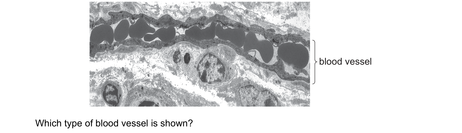
The electron micrograph shows a longitudinal section through a blood vessel. Which type of blood vessel is shown?
Answer: B.
The wall is a single layer of flattened cells. There is no muscle and no elastic tissue, just endothelium and its nucleus bulging into the lumen. Every other vessel on the list has at least a tunica media outside the endothelium, and in a longitudinal section that would show as a thick banded wall.
The red blood cells fill the lumen edge to edge, in single file. They are being squeezed along one behind another rather than flowing in a stream. That only happens in a vessel about the diameter of a red blood cell, roughly 7 µm.
Both of those describe a capillary, which is B.
An arteriole (A) or a venule (D) would be wider, with room for cells to pass side by side, and would have smooth muscle in the wall. A vein (C) would be wider still, with a clearly layered wall and a large lumen holding many cells across.
The two features are not independent, which is worth seeing. The wall is one cell thick and the tube is one cell wide because a capillary exists to exchange materials: a short diffusion distance and a slow, single-file flow are what give the blood time and proximity to swap gases with the tissue.
Question 5
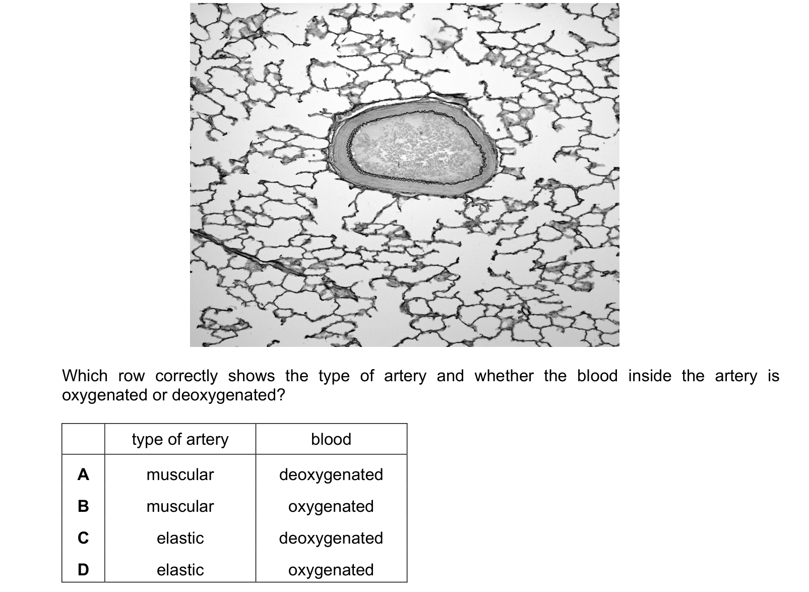
The photomicrograph shows a section through a tissue with an artery. Which row correctly shows the type of artery and whether the blood inside the artery is oxygenated or deoxygenated? Each answer gives, in order: type of artery; blood.
Answer: A.
What is the vessel in? The lacy network filling the rest of the field is alveolar tissue, so this section is from a lung. An artery inside the lung is a branch of the pulmonary artery, and the pulmonary artery is the one artery in the body that carries deoxygenated blood, on its way from the right ventricle to be reoxygenated. That removes B and D.
Which type of artery? Elastic arteries are the largest, right at the heart, where the wall has to absorb the surge of each ventricular contraction. This vessel is well inside the lung, some way from the heart, and its wall is mostly a band of smooth muscle rather than sheets of elastic tissue. It is a muscular artery.
Muscular and deoxygenated, which is A.
The oxygenation half is the memorable part. The usual shortcut, arteries carry oxygenated blood, fails at exactly two vessels: the pulmonary artery, which carries deoxygenated blood away from the heart, and the pulmonary vein, which carries oxygenated blood back to it. The definition that never fails is about direction, not oxygen: an artery carries blood away from the heart.
What this practice covers
These questions are drawn from past CIE 9700 Biology papers and filtered to the circulatory system. You answer, you find out immediately whether you were right, and you get the reasoning for the correct option and for each distractor. Wrong answers go to a mistakes locker so you can come back to exactly those.
Practice is free. You need an account only so your progress and your mistakes are still there next time.
What examiners see students get wrong here
These are the errors that cost marks on the circulatory system, taken from our own topic notes. Read them before you practise and you will recognise the traps in the questions.
- Saying arteries carry oxygenated blood. They carry blood away from the heart; the pulmonary artery is deoxygenated.
- Saying veins have valves "to keep the pressure high". Valves prevent backflow.
- Saying elastic tissue in an artery lets it stretch, and stopping there. The recoil is what maintains flow between heartbeats.
- Saying tissue fluid returns to the capillary because of hydrostatic pressure. It returns by osmosis, because plasma proteins remain behind.
- Saying blood flows slowly in capillaries because they are narrow. It is the large total cross-sectional area of all of them together.
Revise it first
If any of the above is unfamiliar, work through the notes before practising: The circulatory system revision notes.