Testing for biological molecules
Contents: 10 sections
Why these tests turn up so often
Every one of these tests appears in Paper 1 in the same shape: a table of results, and four rows offering different colour changes. The marks are lost on detail rather than on principle. Knowing that Benedict's goes orange is not enough if the question turns on whether the tube was heated, or whether the acid was neutralised first.
So learn each test as a procedure with a reason, not as a colour.
Reducing sugars: the Benedict's test
A reducing sugar has a free aldehyde or ketone group that can donate electrons. All monosaccharides are reducing sugars. Among the disaccharides, maltose and lactose are reducing, and sucrose is not.
Procedure. Add Benedict's solution to the sample and heat in a water bath at about 80 °C for a few minutes.
Result. Blue means no reducing sugar. A positive result runs through green, then yellow, then orange, then brick red as the concentration rises.
The colour change happens because copper(II) ions in the Benedict's solution, which are blue in solution, are reduced to copper(I) oxide, which is an insoluble brick-red precipitate. The sugar is the reducing agent, which is where the name comes from.
Heating is not optional. An unheated tube stays blue whatever is in it, and a question that leaves out the water bath is testing whether you noticed.
Non-reducing sugars
Sucrose gives a negative Benedict's result. To test for it you have to break it into its reducing components first.
- Test a sample with Benedict's. It stays blue, so there is no reducing sugar.
- Take a fresh sample. Add dilute hydrochloric acid and heat. This hydrolyses the glycosidic bond into glucose and fructose.
- Neutralise with sodium hydrogencarbonate or an alkali.
- Now add Benedict's and heat again. An orange or red result means a non-reducing sugar was present.
Step 3 is the one that gets dropped. Benedict's solution is alkaline and simply will not work in an acidic tube, so a candidate who forgets to neutralise reports a false negative. Cambridge asks about this step directly.
The logic of the whole thing is a comparison of two halves of the same sample:
| Sample | First test, Benedict's alone | Second test, after acid and neutralising |
|---|---|---|
| Glucose | orange or red | orange or red |
| Sucrose | blue | orange or red |
| Neither | blue | blue |
A mixture of glucose and sucrose gives a positive first test and a stronger second one.
Starch
Procedure. Add iodine in potassium iodide solution at room temperature. No heating.
Result. Orange-brown stays orange-brown if there is no starch, and turns blue-black if there is.
The colour comes from iodine molecules sitting inside the coiled amylose helix, which is why starch gives the colour and cellulose, which is not coiled, does not.
Lipids: the emulsion test
Procedure. Dissolve the sample in ethanol, shake, then pour the ethanol into water.
Result. A white emulsion forms if lipid is present. A clear solution means none.
Lipid dissolves in ethanol but not in water, so when the ethanol solution meets water the lipid comes out of solution as tiny droplets suspended through the liquid, and those droplets scatter light. The order matters: adding water to the sample before the ethanol gives nothing.
Proteins: the Biuret test
Procedure. Add sodium hydroxide, then a few drops of dilute copper(II) sulfate. No heating.
Result. Blue stays blue if there is no protein. Purple or lilac means peptide bonds are present.
The test detects the peptide bond itself, not the amino acids, so a solution of free amino acids gives a negative result. That distinction is worth holding on to, because it is exactly what a question about the products of protein digestion will turn on.
The tests side by side
| Molecule | Reagent | Heat? | Negative | Positive |
|---|---|---|---|---|
| Reducing sugar | Benedict's | yes | blue | green to brick red |
| Non-reducing sugar | acid, neutralise, then Benedict's | yes | blue | orange or red |
| Starch | iodine in KI | no | orange-brown | blue-black |
| Lipid | ethanol then water | no | clear | white emulsion |
| Protein | Biuret | no | blue | purple |
Two of the five need heat. Reading down that column is the fastest way to eliminate wrong options in a table question.
Semi-quantitative and quantitative measurement
The Benedict's test is semi-quantitative. The colour tells you roughly how much reducing sugar is present, in bands rather than as a number, because more sugar reduces more copper and produces more precipitate.
To turn that into a real measurement you need a colorimeter, which shines light through the sample and measures how much is absorbed or transmitted.
The method is:
- Make up a series of sugar solutions of known concentration.
- Run the Benedict's test on each, filter or centrifuge out the precipitate, and read the remaining blue colour in the colorimeter.
- Plot absorbance against known concentration. This is the calibration curve.
- Treat the unknown sample the same way, read its absorbance, and use the curve to read off its concentration.
Reading the curve the right way
A common trap: as sugar concentration rises, more blue copper(II) is removed, so the remaining solution absorbs less. If the colorimeter is reading the blue supernatant, absorbance falls as concentration rises. If instead the precipitate is left in and the reading is of the red suspension, the relationship runs the other way.
The safe habit is to state what is being measured before deciding which way the line slopes, rather than assuming that more sugar always means a higher reading.
A calibration curve is only valid for the exact conditions used to build it. Change the volume of Benedict's, the heating time or the filter used, and the unknown has to be measured against a fresh curve.
Common mistakes
- Saying "Benedict's turns red" without naming the intermediate colours. A question showing green or yellow is testing the range.
- Forgetting to neutralise after acid hydrolysis in the non-reducing sugar test.
- Heating the Biuret test. It does not need it and the mark scheme does not credit it.
- Saying the Biuret test detects amino acids. It detects the peptide bond.
- Adding water before ethanol in the emulsion test, which gives no emulsion.
Check you have it
Question 1
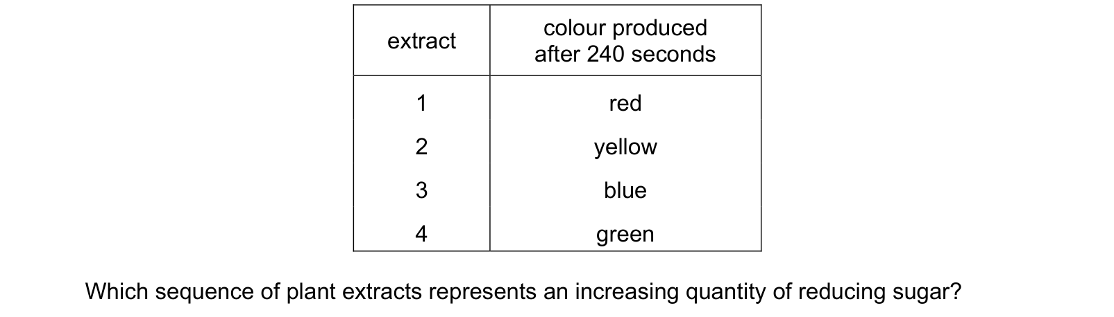
Four extracts from different plant materials were made and tested with Benedict’s solution. The extracts were boiled with Benedict’s solution for 240 seconds and the final colour was recorded. colour produced extract after 240 seconds 1 red 2 yellow 3 blue 4 green Which sequence of plant extracts represents an increasing quantity of reducing sugar?
Answer: D.
blue (none) → green → yellow → orange → brick red (most)
So more reducing sugar means further along that sequence. Reading the table, extract 3 is blue, extract 4 is green, extract 2 is yellow and extract 1 is red.
In order of increasing reducing sugar: 3 → 4 → 2 → 1, which is D.
The colour comes from the reducing sugar donating electrons to the blue copper(II) ions in the Benedict's solution, turning them into a brick-red precipitate of copper(I) oxide. The more sugar there is, the more precipitate forms and the further the colour shifts from blue towards red.
The method matters too. All four extracts were boiled for the same 240 seconds, which is what makes the colours comparable. If one had been heated longer it would have gone further along the sequence for the same amount of sugar, and the comparison would mean nothing. That control is why this counts as a semi-quantitative test rather than just a positive or negative one.
Question 2
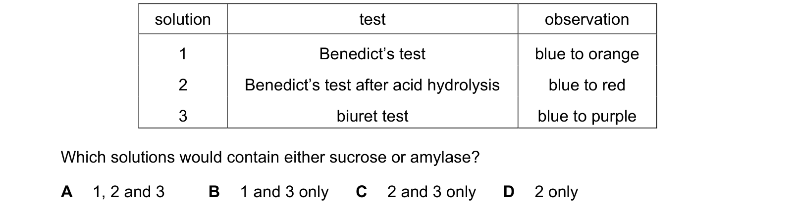
Tests for biological molecules were carried out on three solutions. Each solution contained only one type of biological molecule.
The observations were as follows.
solution test observation
1 Benedict’s test blue to orange
2 Benedict’s test after acid hydrolysis blue to red
3 biuret test blue to purple
Which solutions would contain either sucrose or amylase?
Answer: C.
Solution 1: Benedict's positive without any pretreatment, so it contains a reducing sugar. Sucrose is non-reducing and amylase is a protein, so neither is here.
Solution 2: negative at first, then positive after acid hydrolysis. That is the definition of a non-reducing sugar, and the example asked about is sucrose.
Solution 3: biuret turns purple, which detects peptide bonds, so this is a protein. Amylase is a protein.
So solutions 2 and 3, which is C.
The test that decides it is the biuret one, because it responds to the peptide bond rather than to free amino acids, which is why it identifies an intact enzyme.
Question 3
The concentration of reducing sugar in a solution can be found if an observational measurement is compared to a standard.
Which observational measurement could be used to estimate the concentration of reducing sugar in an unknown solution?
1 the colour of the solution after 20 minutes
2 the time for the first colour change to occur
3 the rate of formation of solid particles
Answer: B.
The Benedict's test is semi-quantitative: more reducing sugar reduces more copper, so more precipitate forms and it forms sooner. Two things can therefore be compared against a standard.
The colour after a fixed time, statement 1, runs further along the blue to brick-red sequence at higher concentrations.
The time to the first colour change, statement 2, is shorter at higher concentrations, which is the version this paper uses elsewhere as a timed end-point.
Statement 3, the rate of formation of solid particles, is not something that can be judged by eye. The precipitate appears as a colour change rather than as countable particles, so it is not an observational measurement.
What the syllabus asks for on this topicSyllabus points
Syllabus points
- Carry out and describe the biochemical tests for reducing sugars, non-reducing sugars, starch, lipids and proteins.
- Describe the use of the Benedict's test as a semi-quantitative measure of reducing sugar concentration.
- Describe the use of a colorimeter and calibration curve to measure concentration.
Related CIE 9700 Biology topics
Not the topic you were looking for? Describe what you are stuck on in your own words and we will take you to the notes that answer it.