The gas exchange system: five questions to try now
Real past-paper questions, the answer key from the mark scheme, and the explanation that goes with it. No account needed to answer them.
Question 1
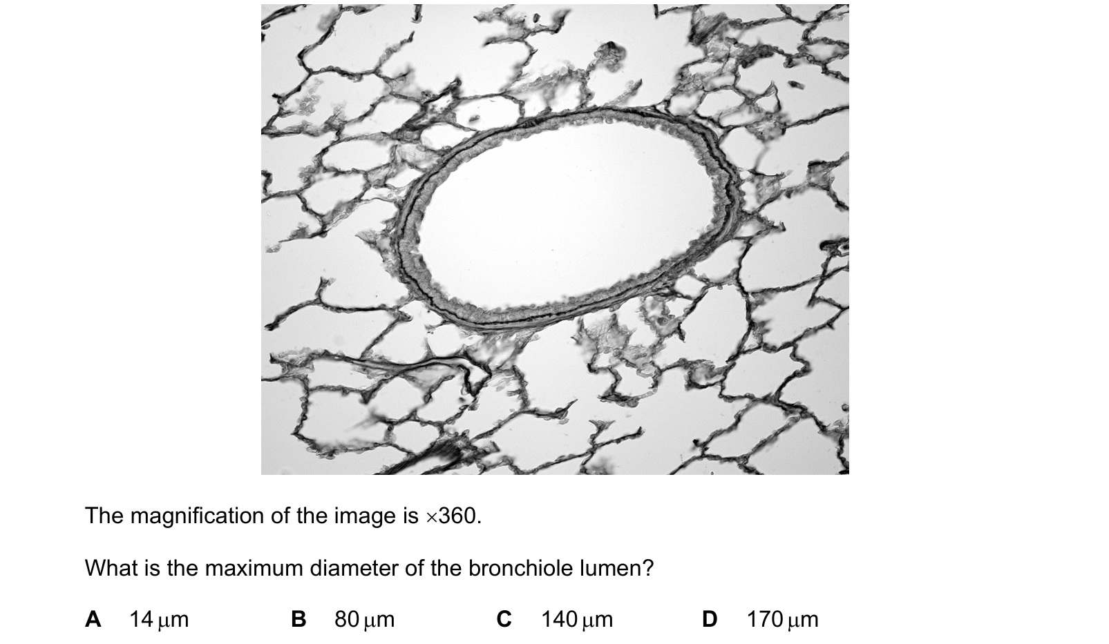
The photomicrograph shows a bronchiole and alveoli.
The magnification of the image is ×360.
What is the maximum diameter of the bronchiole lumen?
Answer: C.
The lumen is the white space inside the ring of epithelium, not the whole tube including its wall. Its longest axis measures about 52 mm on the printed micrograph. So
52 mm = 52 000 µm, and 52 000 ÷ 360 ≈ 144 µm
which is 140 µm to the nearest option, so C.
D, 170 µm, is what you get by measuring the whole bronchiole including its wall rather than just the lumen, which is the trap the word lumen is there to set. B, 80 µm, is roughly the short axis, and the question asks for the maximum diameter. A, 14 µm, is out by a factor of ten.
A check worth making: 140 µm is about a seventh of a millimetre, which is a sensible size for a small airway, and it is far bigger than the alveoli surrounding it. If an answer came out at 14 µm, the bronchiole would be barely twice the width of a capillary and the alveoli around it would have to be smaller still.
Question 2
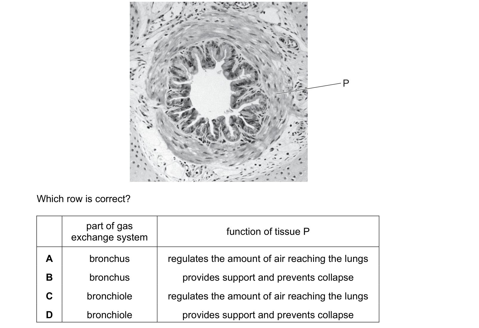
The photomicrograph shows a part of the human gas exchange system with one tissue labelled P. Which row is correct? Each answer gives, in order: part of gas exchange system; function of tissue P.
Answer: C.
Which part of the system? The airway has a folded epithelium and a distinct ring of muscle around it, and no cartilage anywhere in the wall. No cartilage means it cannot be a bronchus or the trachea, so it is a bronchiole. That removes A and B.
What does P do? P points at the band around the outside of the epithelium, which is smooth muscle. Smooth muscle contracts and relaxes to change the diameter of the bronchiole, so it regulates the amount of air reaching the lungs. That is C.
D offers support and prevention of collapse, which is the job of cartilage, and the absence of cartilage is precisely what identified this as a bronchiole in the first place. So D contradicts its own first column, which is a useful thing to notice: in a two-column row, the columns have to be consistent with each other as well as with the picture.
The division of labour is worth holding on to. Bigger airways need to stay permanently open, so they have cartilage. Smaller ones need to be adjustable, so they have muscle instead, and that is what makes bronchoconstriction possible in asthma.
Question 3
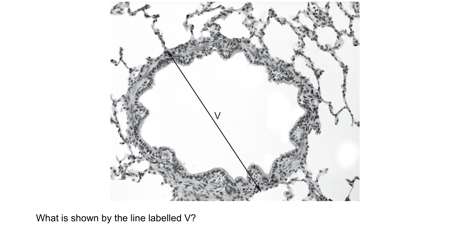
The photomicrograph shows a cross-section of part of the gas exchange system of a mammal. What is shown by the line labelled V?
Answer: B.
It is a large tube lined with epithelium, surrounded on all sides by the lacy network of alveoli, and there is no cartilage anywhere in its wall. An airway inside the lung tissue with no cartilage is a bronchiole, and V runs right across its lumen. That is B.
Each wrong answer fails on scale or on setting.
A, an alveolus. The alveoli are the small, thin-walled spaces making up the network around the tube, an order of magnitude smaller than the structure V spans.
C, a capillary. A capillary is about 7 µm across, just wide enough for one red blood cell. At this magnification it would be a hairline, not the widest thing in the field.
D, a trachea. The trachea is a single tube in the neck, held open by C-shaped rings of cartilage, and it is not embedded in lung tissue. Seeing alveoli all round the outside rules it out on its own.
The absence of cartilage is the feature to look for first in any airway section. Trachea and bronchi have it; bronchioles do not, and rely on smooth muscle instead.
Question 4
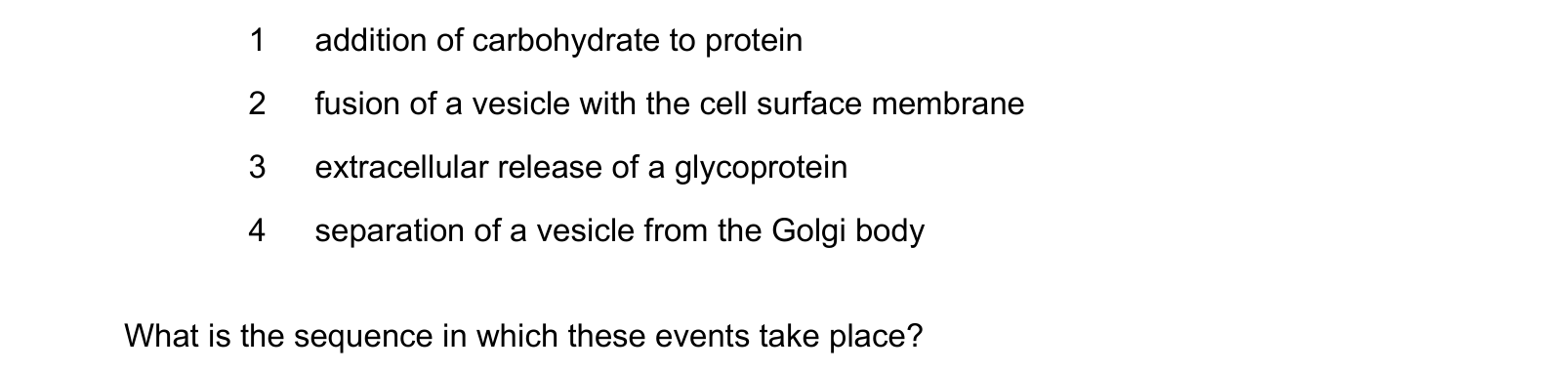
When mucus is secreted from a goblet cell, these events take place. What is the sequence in which these events take place?
Answer: A.
1 first: addition of carbohydrate to protein. This happens in the Golgi body, and it is what turns the protein into a glycoprotein in the first place.
4 next: separation of a vesicle from the Golgi body. Only once the molecule is finished is it packaged and pinched off.
2 then: fusion of a vesicle with the cell surface membrane. The vesicle travels out and its membrane joins the cell surface membrane.
3 last: extracellular release of a glycoprotein. Fusing is what opens the vesicle to the outside, so the release cannot come before it.
1 → 4 → 2 → 3, which is A.
C and D both start with 4, taking a vesicle from the Golgi before anything has been done to the protein inside it. B and D put 3 before 2, releasing the mucus outside the cell before the vesicle has reached the membrane, which is the step that makes release possible.
That whole final sequence, 4 then 2 then 3, is exocytosis, and it costs ATP: the vesicle is walked along the cytoskeleton rather than drifting.
Question 5
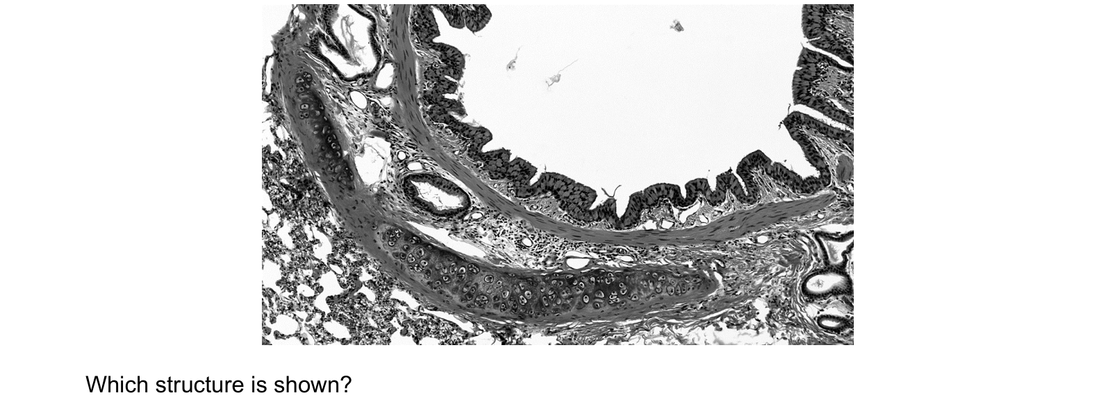
The photomicrograph shows a section through part of the human gas exchange system.
Which structure is shown?
Answer: C.
The lumen is lined with a folded ciliated epithelium, then there is a band of smooth muscle, and then the feature that decides it: a thick pale plate of cartilage, recognisable by the cells sitting in small spaces within it. Around the outside, bottom left, is the lacy network of alveoli.
Cartilage present, and lung tissue all around it, is a bronchus, which is C.
D, the trachea, also has cartilage, but as complete C-shaped rings, and it runs down the neck rather than through the lung. Alveoli in the same section rule it out.
B, a bronchiole, has no cartilage at all. That is its defining absence.
A, an alveolus, is one of the small thin-walled spaces in the network at the edge of the picture, far smaller than the structure filling the field.
The useful sequence for any airway section: look for cartilage first, because it splits the airways in two. If it is there, you are in the trachea or a bronchus and the question is which. If it is not, you are in a bronchiole.
What this practice covers
These questions are drawn from past CIE 9700 Biology papers and filtered to the gas exchange system. You answer, you find out immediately whether you were right, and you get the reasoning for the correct option and for each distractor. Wrong answers go to a mistakes locker so you can come back to exactly those.
Practice is free. You need an account only so your progress and your mistakes are still there next time.
What examiners see students get wrong here
These are the errors that cost marks on the gas exchange system, taken from our own topic notes. Read them before you practise and you will recognise the traps in the questions.
- Saying bronchioles contain cartilage. They do not, and this is the single most tested fact in the topic.
- Saying alveoli have goblet cells or cilia. They have neither, because both would be too thick.
- Saying the alveolar wall is "thin" without saying it is one cell thick.
- Saying a large surface area alone explains fast exchange. The short distance and the maintained gradient matter just as much.
- Saying ventilation maintains the gradient, and stopping there. The blood supply is the other half.
Revise it first
If any of the above is unfamiliar, work through the notes before practising: The gas exchange system revision notes.