The heart: five questions to try now
Real past-paper questions, the answer key from the mark scheme, and the explanation that goes with it. No account needed to answer them.
Question 1
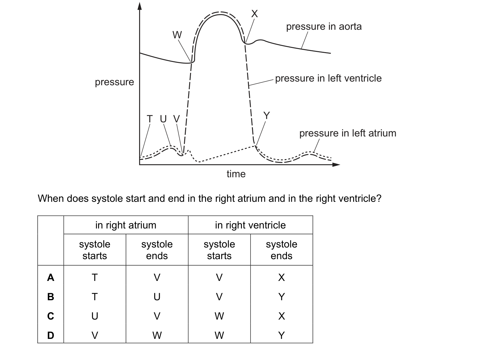
The graph shows some blood pressure changes that occur in a human during one cardiac cycle. When does systole start and end in the right atrium and in the right ventricle?
Answer: A.
Atrial systole starts at T, where the atrial pressure begins to climb, and it ends at V, where that pressure has fallen back and the ventricle takes over. U is only the peak of the atrial wave, part way through the contraction, not the end of it.
Ventricular systole starts at V, where the ventricular line begins its steep rise, and ends at X, where that line falls back to meet the aorta and the semilunar valve shuts.
T, V, V, X, which is A.
Y is later than the end of systole. It is where the ventricular pressure has dropped all the way to the atrial pressure and the atrioventricular valve reopens. Between X and Y the ventricle is already relaxing, so that stretch belongs to diastole, and B and D both put the end of systole there.
W is where the semilunar valve opens, in the middle of ventricular systole rather than at its start, which is what makes C wrong.
One moment doing two jobs is the feature to hold on to: V ends atrial systole and starts ventricular systole, which is why it appears twice in the correct row.
Question 2
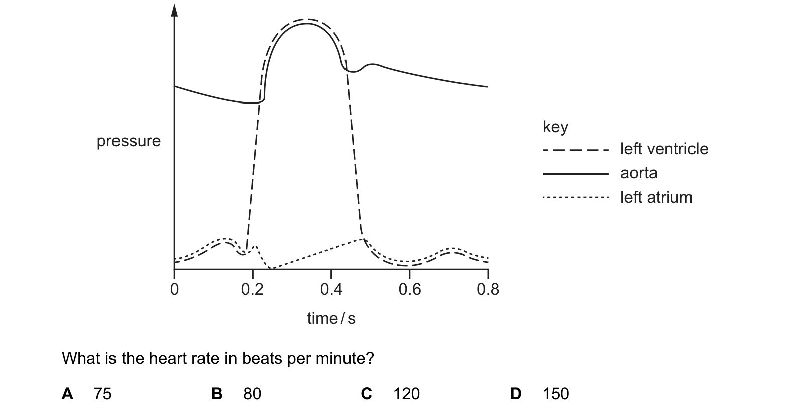
The graph shows the changes in pressure that occur in the left side of the heart during one cardiac cycle. pressure time / s
What is the heart rate in beats per minute?
Answer: A.
The graph shows exactly one complete cycle across its full width. At 0.8 s the aortic pressure has fallen back to where it was at 0 s and is still declining, the ventricle is back at rest, and the whole pattern is about to repeat. So one cardiac cycle takes 0.8 s.
60 ÷ 0.8 = 75 beats per minute, which is A.
Each wrong answer is a different cycle length read off the axis. 80 comes from 0.75 s, 120 from 0.5 s, which is the width of the ventricular pressure peak rather than the whole beat, and 150 from 0.4 s.
That is the mistake to guard against: the ventricle only spends part of the cycle contracting, so the width of the tall dashed peak is systole, not the beat. The beat runs from the start of one atrial contraction to the start of the next, and includes the long quiet stretch of diastole afterwards.
75 beats per minute is also a sensible resting rate, which is worth using as a check. An answer of 150 would be someone sprinting.
Question 3
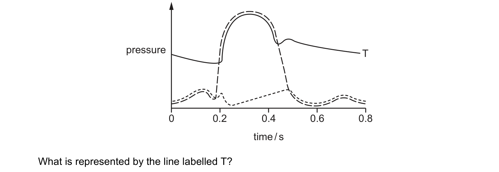
The graph shows the pressure changes in three structures of the right side of the heart during the cardiac cycle. time / s
What is represented by the line labelled T?
Answer: A.
T stays high throughout. It never falls near zero, and after the peak it does not drop back with the others but settles to a slow decline. That is a blood vessel, not a chamber: the artery keeps its pressure between beats because its elastic walls recoil onto the blood. So T is the pulmonary artery, which is A.
The small notch in T just after the peak clinches it. That is the dicrotic notch, the moment the semilunar valve snaps shut and blood rebounds against it. It appears in an artery trace and nowhere else.
B and C are the other two lines. The one that rises from near zero to the peak and falls straight back is the right ventricle, and the low line with small waves is the right atrium. Both empty, so both fall to almost nothing.
D, the vena cava, would be lower still and almost flat. Nothing on the graph both peaks with the ventricle and stays high afterwards except the artery leaving it.
Question 4
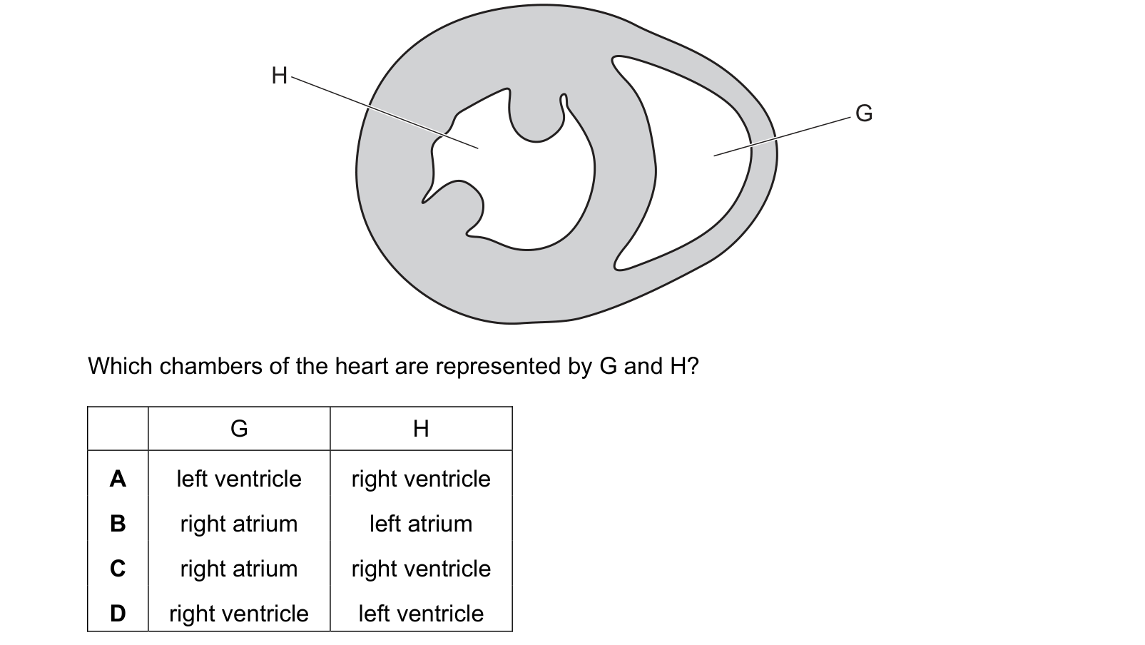
The diagram shows a cross-section through a mammalian heart. Which chambers of the heart are represented by G and H? Each answer gives, in order: G; H.
Answer: D.
Now tell the two apart by the wall. H is the large, roughly circular chamber surrounded by a very thick band of muscle, so H is the left ventricle. It has to generate enough pressure to drive blood round the entire body, and the circular shape is what lets a thick wall squeeze evenly.
G is the crescent-shaped chamber wrapped round the outside of it, with a much thinner wall, so G is the right ventricle. It only pumps as far as the lungs, which are close by and easily damaged by high pressure, so it needs perhaps a third of the muscle.
G right, H left, which is D. A has them the correct way round in kind but the wrong way round in fact.
The crescent shape is the giveaway worth remembering. The right ventricle is not a separate tube alongside the left one; it wraps around it, so in cross-section it always appears as a curved slit against a circle.
Question 5
The diagram shows pressure changes in the left side of the heart during the cardiac cycle. time / s
What happens in the heart at X?
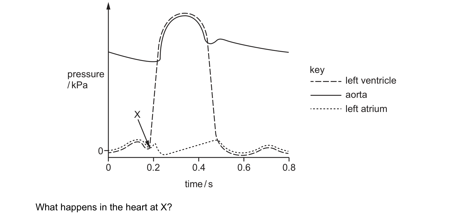
Answer: A.
A valve is held open by the pressure behind it and shut by the pressure in front. The moment the ventricle's pressure exceeds the atrium's, blood tries to flow backwards, the cusps are pushed together, and the atrioventricular valves close. That is A, and it is the start of ventricular systole.
D happens next but not yet. The semilunar valves cannot open until the ventricle beats the pressure in the aorta, which is much higher than the atrium's. On this graph that is where the dashed line meets the solid aorta line, further up and slightly later.
C is the mirror image of D, at the end of systole, where the falling ventricle line rejoins the aorta line.
B is the last event of the cycle, where the ventricle line falls back below the atrium line at about 0.48 s.
The short gap between X and the semilunar valves opening is isovolumetric contraction: all four valves shut, no blood moving, and the pressure climbing almost vertically.
What this practice covers
These questions are drawn from past CIE 9700 Biology papers and filtered to the heart. You answer, you find out immediately whether you were right, and you get the reasoning for the correct option and for each distractor. Wrong answers go to a mistakes locker so you can come back to exactly those.
Practice is free. You need an account only so your progress and your mistakes are still there next time.
What examiners see students get wrong here
These are the errors that cost marks on the heart, taken from our own topic notes. Read them before you practise and you will recognise the traps in the questions.
- Saying the left ventricle is thicker "because it pumps more blood". Both ventricles pump the same volume; the left generates more pressure.
- Saying valves open and close by muscle action. They are opened and closed by pressure differences.
- Saying the tendinous cords open the valves. They prevent them inverting.
- Saying the SAN sends a nerve impulse. It is cardiac muscle, and the wave is electrical excitation spreading through muscle.
- Forgetting why the AVN delays. Without it, the atria and ventricles would contract together and the ventricles would fill poorly.
Revise it first
If any of the above is unfamiliar, work through the notes before practising: The heart revision notes.