The microscope in cell studies: five questions to try now
Real past-paper questions, the answer key from the mark scheme, and the explanation that goes with it. No account needed to answer them.
Question 1
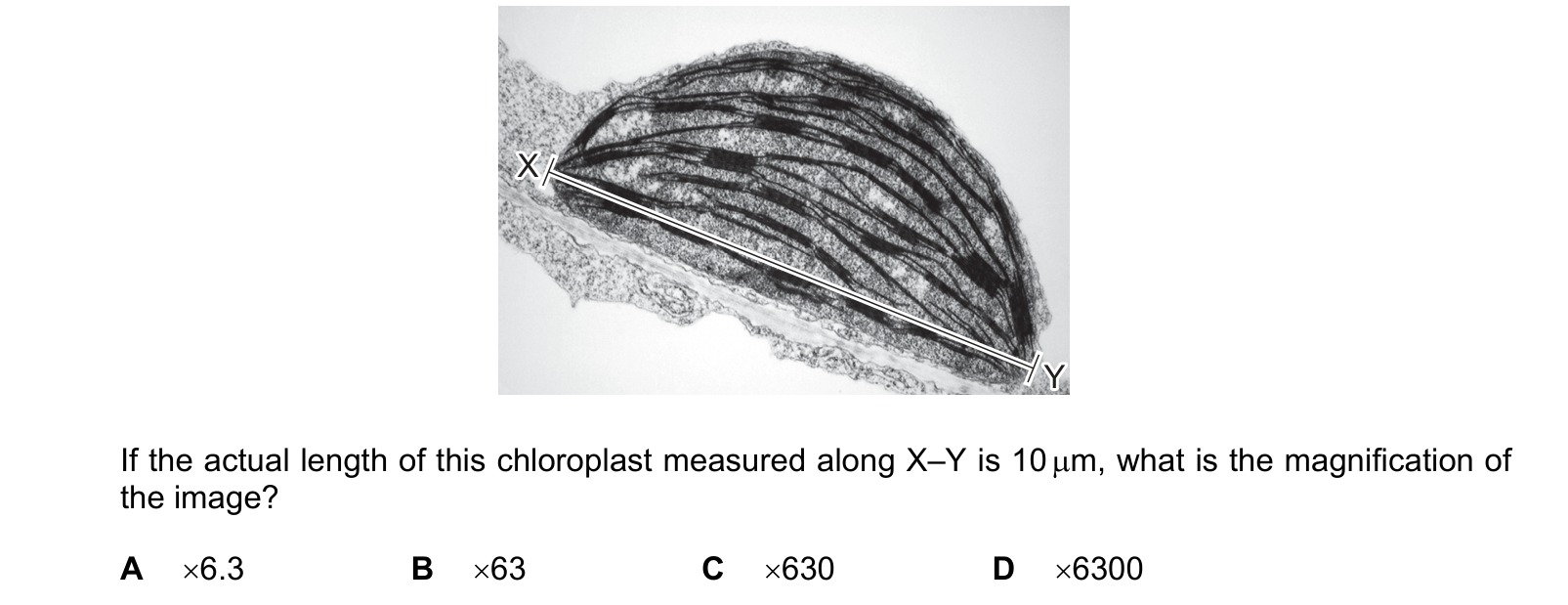
The electron micrograph shows a chloroplast from a tobacco leaf. If the actual length of this chloroplast measured along X–Y is 10 µm, what is the magnification of the image?
Answer: D.
Measured with a ruler on the printed page, the line from X to Y is about 63 mm. The actual length is 10 µm.
Convert the image length to micrometres: 1 mm = 1000 µm, so
63 mm = 63 000 µm
magnification = 63 000 / 10 = × 6300
which is D.
B, × 63, is the answer with the conversion left out, dividing 63 mm by 10 µm as though the units matched. That single step is worth three of the four options, since A and C are the same slip with the decimal point moved once more.
The safest routine is to convert both measurements to micrometres before dividing, and to notice that the magnification has no unit at all: it is a ratio of two lengths, so if a unit survives into the answer something has gone wrong.
A magnification of several thousand also makes sense for an electron micrograph. A light microscope reaches about × 1500 at best, so anything in the thousands or above must have come from an electron microscope, which is what the stem says this is.
Question 2
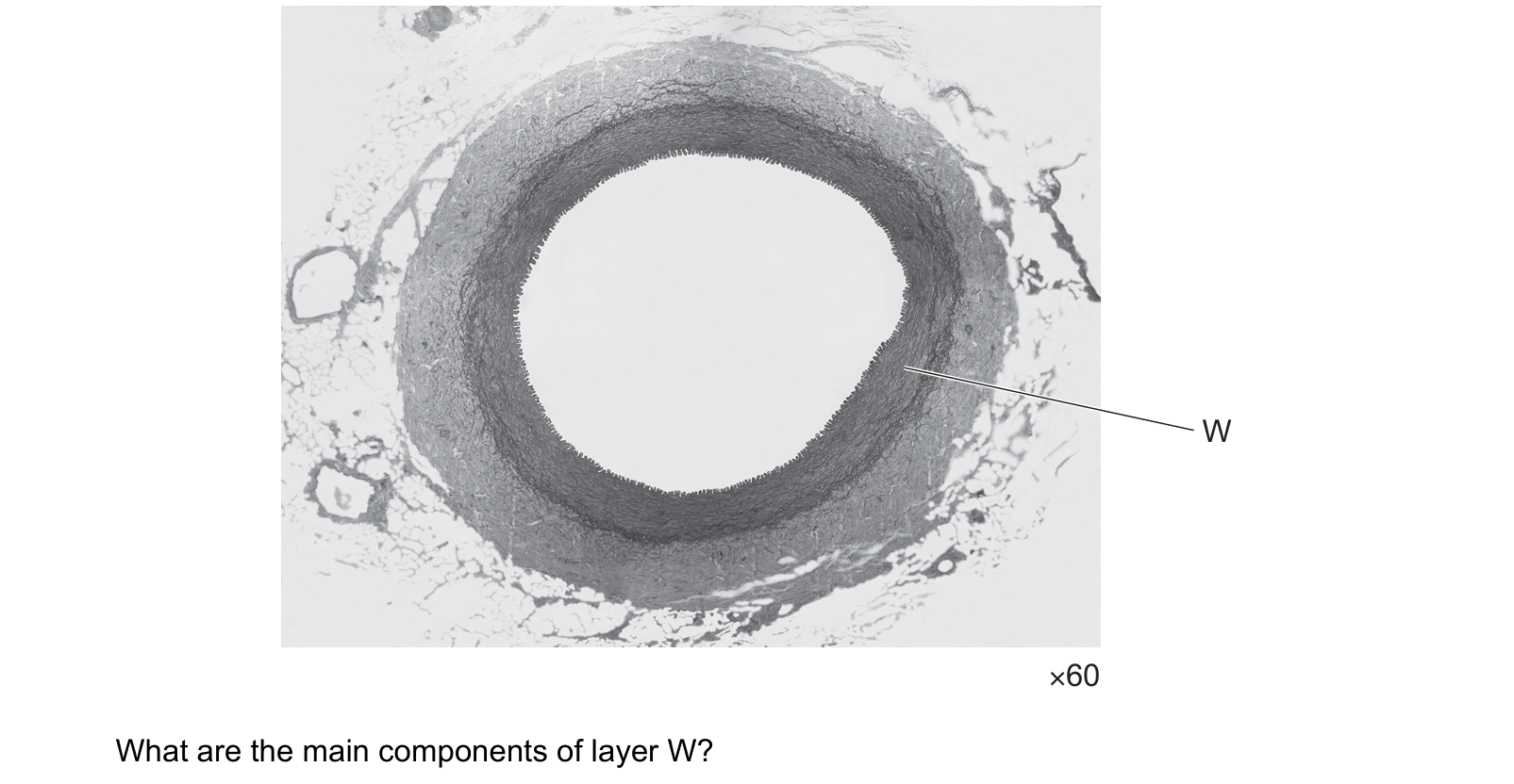
The photomicrograph shows a section through a structure found in mammals viewed using a light microscope. What are the main components of layer W?
Answer: C.
The tunica media is built from smooth muscle and elastic fibres, which is C. The muscle contracts to narrow the lumen and control how much blood reaches a tissue; the elastic fibres stretch as blood is forced in and recoil between beats.
The proportions also tell you which vessel this is. The wall is very thick compared with the lumen and the lumen is held round rather than collapsed, which is an artery. A vein at the same magnification has a thin wall and a wide, often flattened lumen.
D describes the wrong layer. Squamous epithelial cells forming an endothelium are the innermost lining, a single cell thick, which is the thin dark line right at the edge of the lumen rather than the broad band W points to.
A and B leave out the smooth muscle. Collagen is certainly present, mostly in the outer tunica externa, where its job is to stop the vessel bursting, but naming collagen alone or collagen with elastic fibres misses the layer's defining component.
Question 3
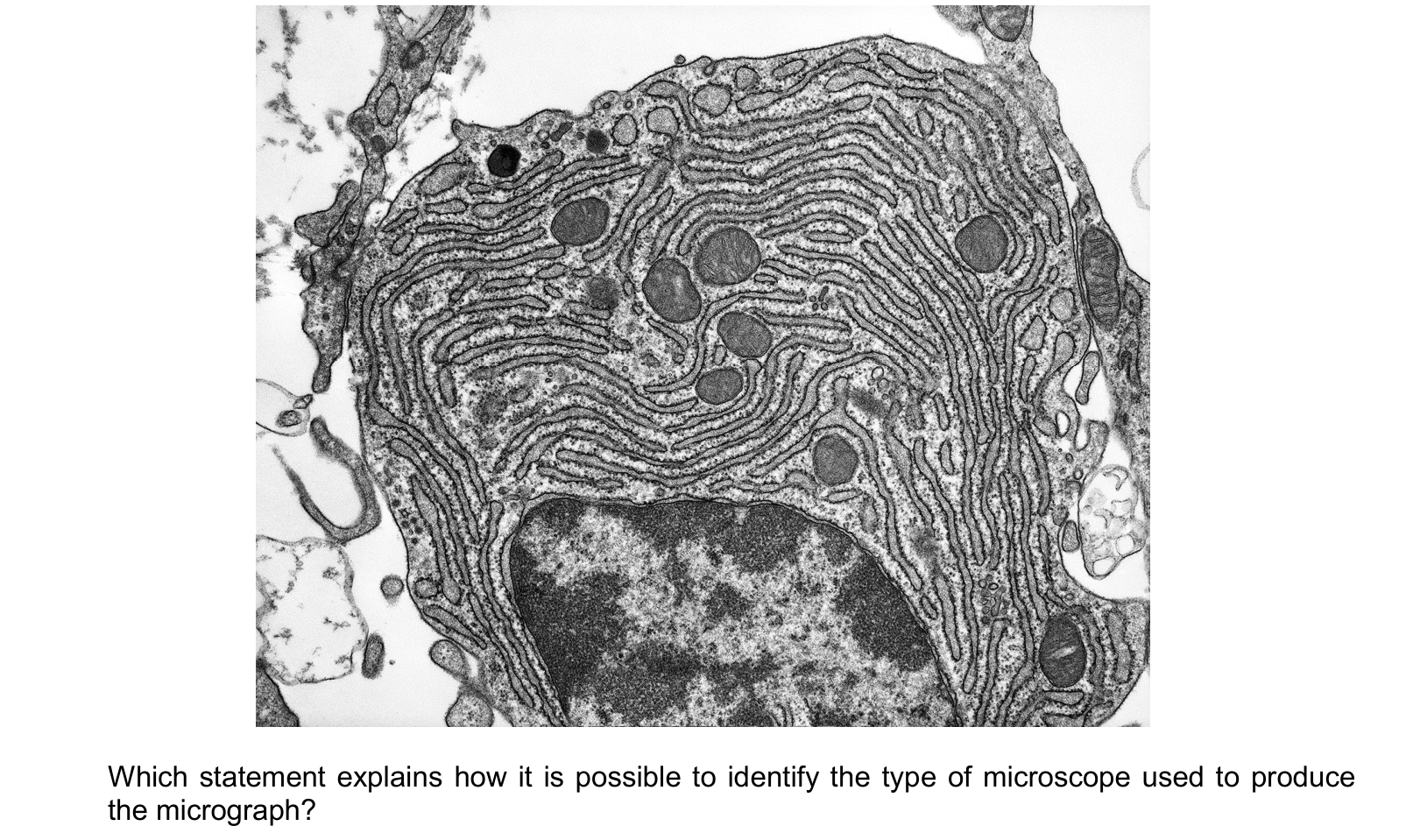
A cell is shown in the micrograph.
Which statement explains how it is possible to identify the type of microscope used to produce the micrograph?
Answer: D.
The membranes of the endoplasmic reticulum in this micrograph are visibly studded with ribosomes, the fine dark granules along each cisterna. A ribosome is about 20 nm across, and a light microscope cannot resolve anything smaller than roughly 200 nm, whatever its magnification. Seeing ribosomes at all is therefore only possible with an electron microscope. That is D.
A has the right microscope for the wrong reason. A nucleus is around 10 µm and is perfectly visible with a light microscope; it is one of the first things a student sees in an onion cell. So a visible nucleus proves nothing.
B is contradicted by the image. The endoplasmic reticulum is not just visible, it fills most of the cell.
C fails twice. There are no chloroplasts here, and chloroplasts are large enough to be seen with a light microscope anyway, so they could not distinguish the two.
The idea being tested is resolution, not magnification. Magnification makes an image bigger; resolution decides whether two things close together stay separate. That is why the limit is set by the wavelength used, and why electrons, with a far shorter wavelength than light, can show a ribosome at all.
Question 4
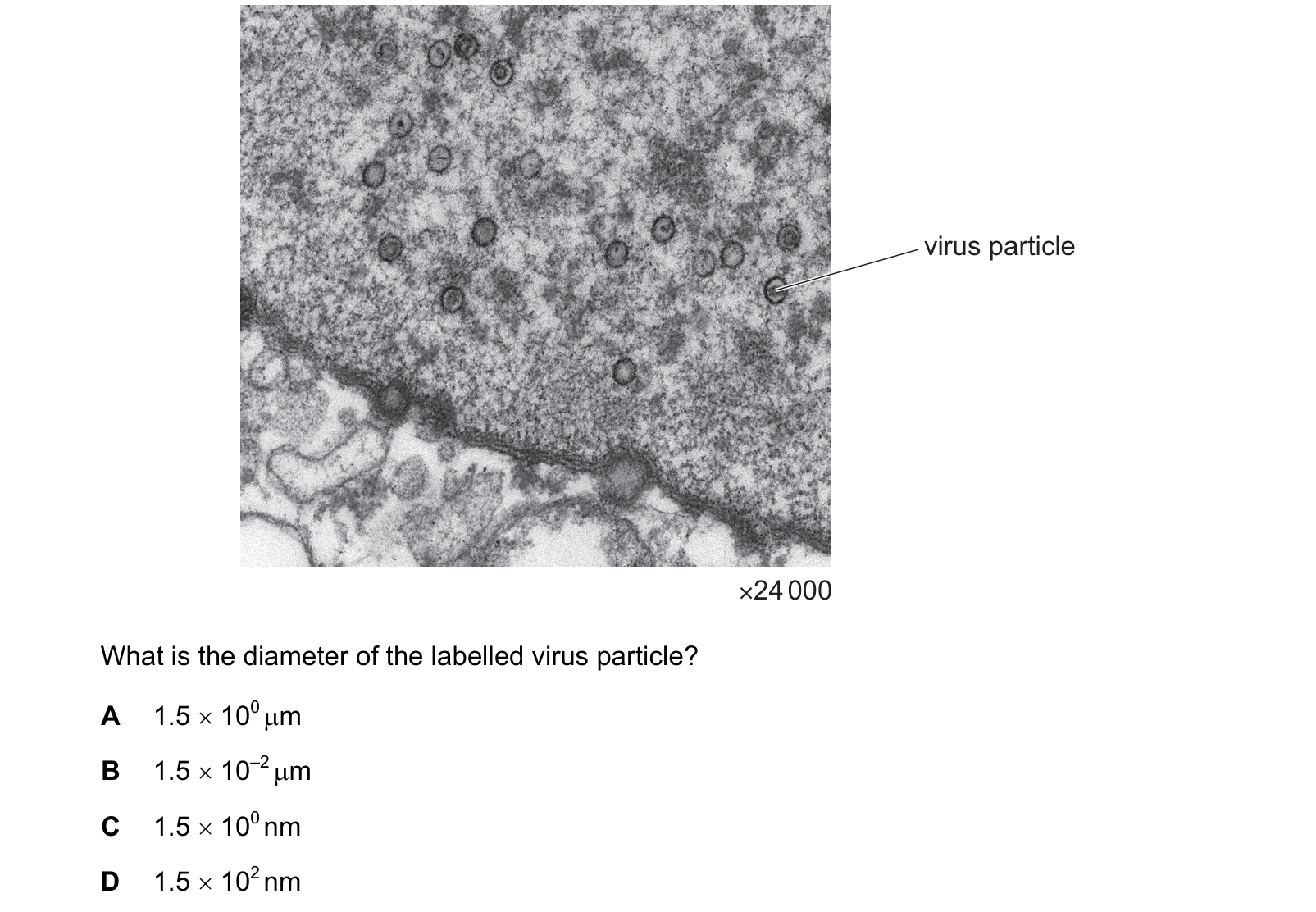
The diagram shows an electron micrograph of virus particles in a human nucleus. What is the diameter of the labelled virus particle?
Answer: D.
Measure the labelled particle on the micrograph and it is about 3.6 mm across. So
3.6 mm ÷ 24 000 = 1.5 × 10⁻⁴ mm
Now convert. 1.5 × 10⁻⁴ mm is 1.5 × 10⁻¹ µm, which is 1.5 × 10² nm, or 150 nm. That is D.
The four options are the same number with four different combinations of power and unit, so the whole question is the conversion. Write them all in nanometres and they separate at once: A is 1500 nm, B is 15 nm, C is 1.5 nm, D is 150 nm.
Each wrong one is also wrong biologically, which is the check to make if you are unsure of the arithmetic. 1.5 nm is the width of a DNA double helix. 15 nm is about the size of a single protein. 1.5 µm is the length of a small bacterium, and the micrograph shows dozens of these particles fitting inside one nucleus. A virus of 100 to 200 nm is what the picture is consistent with.
Question 5
The photomicrograph is of a plant cell. The cell is 25 µm in width from X to Y. What is the magnification of the photomicrograph?
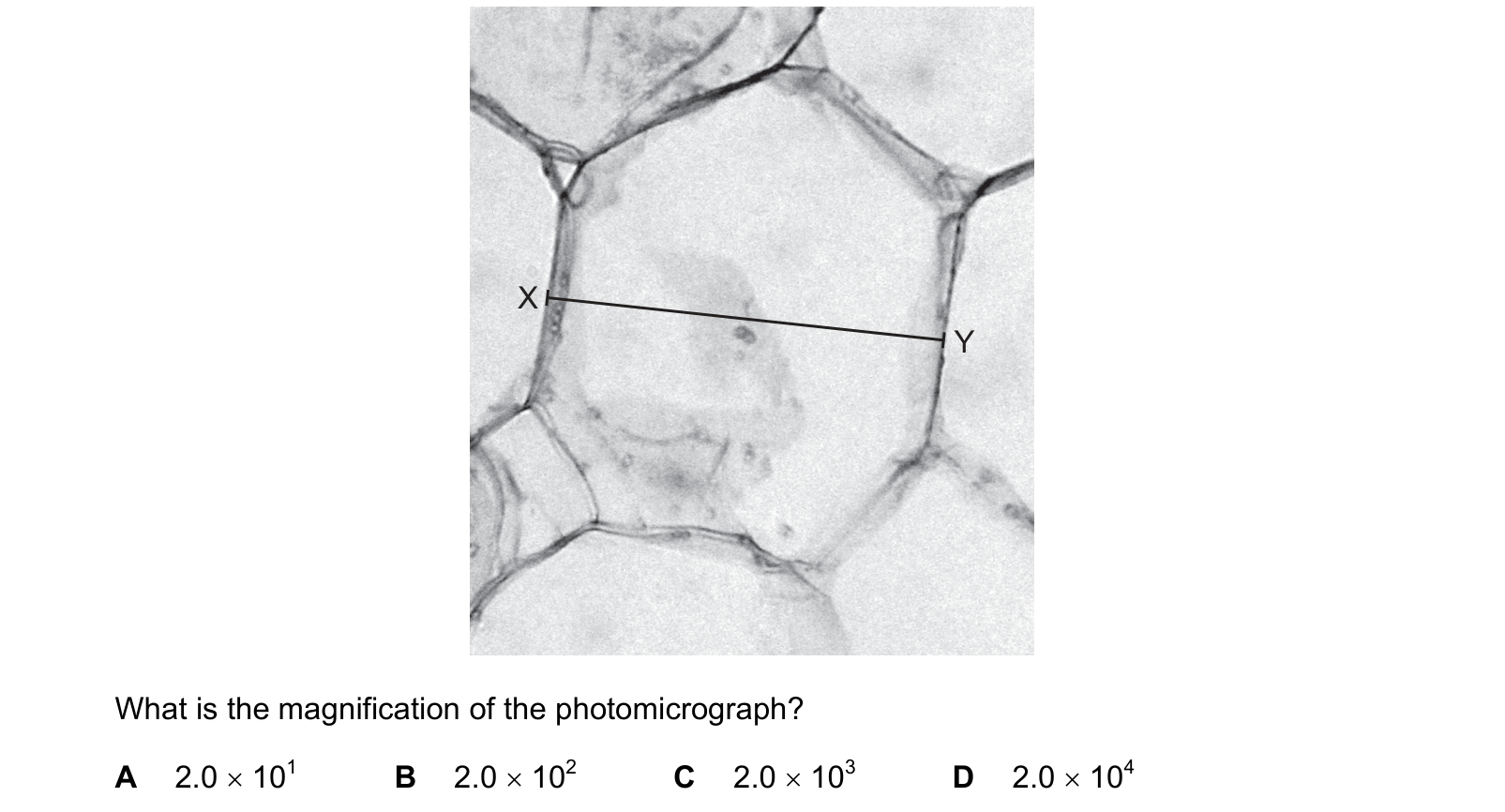
Answer: C.
Measure the line from X to Y on the photomicrograph and it is about 50 mm. The stem tells you the cell is 25 µm wide between those points. Put the image size into micrometres:
50 mm = 50 000 µm
magnification = 50 000 ÷ 25 = 2000, which is 2.0 × 10³, so C.
The four options are decades apart, which tells you the examiner is testing the unit conversion rather than the division. Forget to convert millimetres to micrometres and you get 2, which is not offered; convert by the wrong factor and you land on one of the neighbours.
A sanity check that costs nothing: this is a light microscope image of a plant cell, and light microscopes top out at around ×1500 for a useful image, with printed photomicrographs commonly a couple of thousand. 2.0 × 10⁴ would be electron microscope territory, and at 2.0 × 10¹ a 25 µm cell would be half a millimetre across on the page, far too small to see the walls in this detail.
What this practice covers
These questions are drawn from past CIE 9700 Biology papers and filtered to the microscope in cell studies. You answer, you find out immediately whether you were right, and you get the reasoning for the correct option and for each distractor. Wrong answers go to a mistakes locker so you can come back to exactly those.
Practice is free. You need an account only so your progress and your mistakes are still there next time.
What examiners see students get wrong here
These are the errors that cost marks on the microscope in cell studies, taken from our own topic notes. Read them before you practise and you will recognise the traps in the questions.
- Giving magnification a unit. It is a ratio and has none.
- Doing the calculation without converting both measurements to the same unit first.
- Saying an electron microscope "magnifies more", when the point is that it resolves more. Higher magnification without higher resolution shows nothing new.
- Claiming an electron microscope shows living specimens.
- Forgetting to recalibrate the graticule after changing the objective lens.
- Confusing a TEM image with an SEM image: internal detail means transmission.
Revise it first
If any of the above is unfamiliar, work through the notes before practising: The microscope in cell studies revision notes.