Gas exchange in humans
Contents: 8 sections
Features of a gas exchange surface
Every gas exchange surface in biology, from an alveolus to a gill to a leaf, has the same four features, and each has the same reason:
- Large surface area — more gas can diffuse at once.
- Thin, usually one cell thick — a short diffusion distance.
- Good blood supply — carries gases away, keeping the concentration gradient steep.
- Well ventilated — brings fresh air in, keeping the gradient steep.
The last two are the ones commonly left out. Both work by maintaining the concentration gradient, and saying so is what earns the mark.
The alveoli deliver all four: there are about 300 million of them giving a surface area of roughly 70 m², their walls are one cell thick, each is wrapped in capillaries, and breathing refreshes the air inside.
The breathing system
Air travels: nose or mouth → trachea → bronchi → bronchioles → alveoli.
- Trachea — held open by rings of cartilage, so it cannot collapse when pressure inside drops during inhalation. The rings are C-shaped so the oesophagus behind can expand when swallowing.
- Bronchi — one to each lung, also supported by cartilage.
- Bronchioles — finer tubes, no cartilage, with muscle in their walls that can narrow them.
- Alveoli — tiny air sacs where gas exchange happens.
Gas exchange at the alveolus
Air in the alveolus is high in oxygen and low in carbon dioxide. Blood arriving in the capillary is the reverse. So:
- Oxygen diffuses from alveolus into blood, and combines with haemoglobin in red blood cells.
- Carbon dioxide diffuses from blood into the alveolus and is breathed out.
Both are diffusion, down concentration gradients, and neither needs energy.
Inspired and expired air
| Inspired | Expired | |
|---|---|---|
| Oxygen | 21% | 16% |
| Carbon dioxide | 0.04% | 4% |
| Nitrogen | 78% | 78% |
| Water vapour | variable | saturated |
| Temperature | ambient | warmer, at body temperature |
Two points are often got wrong. Expired air still contains plenty of oxygen, about 16%, which is why mouth-to-mouth resuscitation works. And nitrogen does not change, because it takes no part in respiration.
The test for carbon dioxide is limewater, which turns from clear to milky or cloudy. In the standard experiment, breathing in through one tube and out through another shows expired air turns limewater milky much faster than inspired air does.
Hydrogencarbonate indicator is the other reagent worth knowing. It is orange in normal air, turns yellow when carbon dioxide increases, and purple or magenta when carbon dioxide falls. It is used to show respiration and photosynthesis in the same experiment.
Ventilation
Air moves because of pressure differences, and pressure changes because volume changes. Getting the causal order right is what these questions test.
Breathing in:
- External intercostal muscles contract, pulling the ribcage up and out.
- The diaphragm contracts and flattens, moving down.
- Volume of the thorax increases.
- Pressure inside decreases, falling below atmospheric pressure.
- Air flows in.
Breathing out:
- The intercostal muscles relax, so the ribcage moves down and in.
- The diaphragm relaxes and returns to its domed shape.
- Volume decreases.
- Pressure increases above atmospheric.
- Air flows out.
Note that the diaphragm contracts to flatten and relaxes to dome. Answers frequently reverse this. And air is never pulled or pushed by the lungs themselves: they have no muscle, and simply follow the movement of the chest wall.
Physical activity
During exercise, muscles respire faster, using more oxygen and producing more carbon dioxide.
Increased carbon dioxide in the blood is detected by the brain, which increases the rate and depth of breathing. Deeper breaths bring in more air per breath, and faster breathing brings in more breaths, so more oxygen is delivered and carbon dioxide removed more quickly.
Breathing stays raised after exercise stops, to repay the oxygen debt from anaerobic respiration and to remove the lactic acid that built up.
Protecting the lungs
- Goblet cells produce mucus, which traps dust, bacteria and other particles.
- Ciliated cells have cilia that beat in waves, sweeping the mucus up the airways to the throat, where it is swallowed.
Smoking damages this system. Tar paralyses and eventually destroys the cilia, so mucus is no longer swept away. It collects in the airways, and the only way to shift it is coughing, which is smoker's cough. The trapped bacteria in the stationary mucus make chest infections and bronchitis much more likely.
Check you have it
Question 1
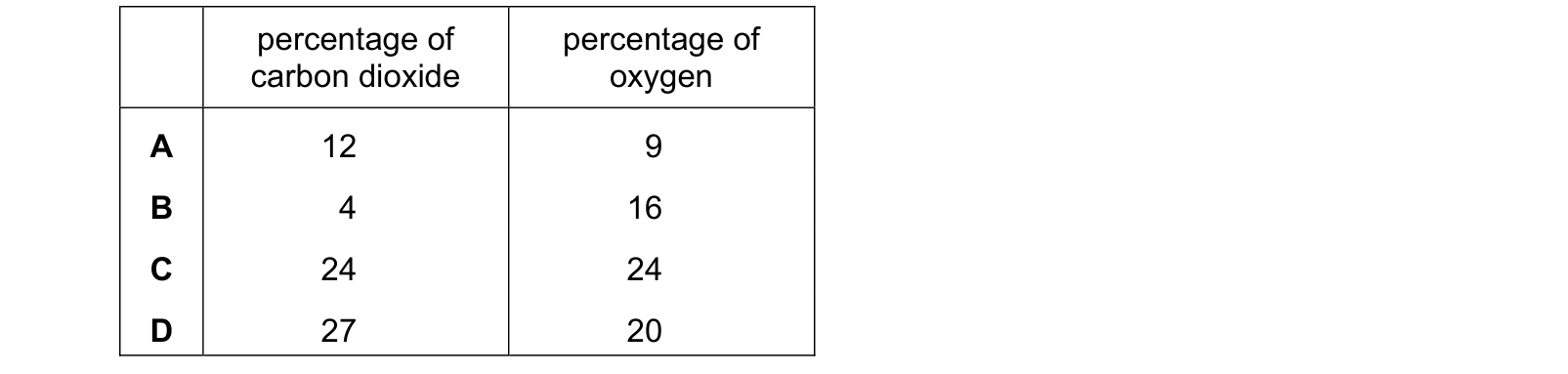
Which row shows the approximate percentage of gases in expired air? Each answer gives, in order: percentage of carbon dioxide; percentage of oxygen.
Answer: B.
Check the order of the columns, since this row asks for carbon dioxide first. Respiration uses oxygen and produces carbon dioxide, so relative to inspired air, oxygen falls from 21% to 16% and carbon dioxide rises from 0.04% to 4%.
The wrong rows are all implausible on the totals as well as the biology. 24% and 24% (C) and 27% and 20% (D) would each require expired air to be nearly half carbon dioxide and oxygen combined, when about 78% of it is nitrogen, which is neither used nor produced.
12% and 9% (A) removes far more oxygen than the body takes on a single pass.
What is worth noticing is how little changes. Only about a quarter of the inhaled oxygen is used, which is why exhaled air still supports mouth-to-mouth resuscitation.
Question 2
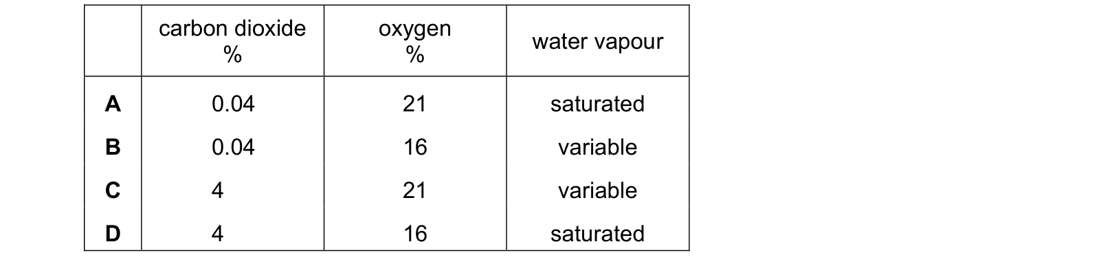
Which row shows the composition of expired air from a healthy person? Each answer gives, in order: carbon dioxide %; oxygen %; water vapour.
Answer: D.
All three follow from what happens in the lungs. Respiration uses oxygen, so it falls from 21% to 16%, and produces carbon dioxide, so it rises from 0.04% to 4%. And air passing over the moist surfaces of the airways and alveoli picks up water until it is close to saturated.
The wrong rows each keep an inspired value. 0.04% carbon dioxide and 21% oxygen are both inspired figures, so a row containing either has not changed the gas that should have changed.
Variable describes the water content of inspired air, which depends entirely on the weather. Expired air is saturated whatever the conditions outside, which is why breath mists on a cold day regardless of humidity.
Expired air is also warmer, having been at body temperature.
Question 3
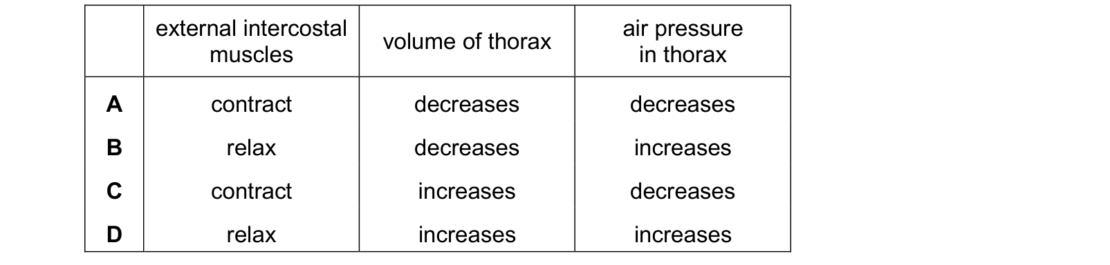
What causes air to enter the lungs? Each answer gives, in order: external intercostal muscles; volume of thorax; air pressure in thorax.
Answer: C.
The three columns are a chain of cause and effect, so they have to agree.
The external intercostals contract, pulling the ribs up and out. Together with the diaphragm flattening, that increases the volume of the thorax. The same amount of air in a larger space means a lower pressure, and air then flows in from the higher pressure outside.
Every wrong row breaks the chain somewhere. Contracting the external intercostals cannot decrease the volume, and an increased volume cannot give an increased pressure.
The row with the muscles relaxing describes breathing out, where the volume falls and the pressure rises.
The lungs have no muscle of their own, so they never draw air in. They are inflated by the pressure difference the ribs and diaphragm create around them.
What the syllabus asks for on this topicSyllabus points
Syllabus points
- Describe the features of gas exchange surfaces.
- Identify the parts of the breathing system and state their functions.
- Compare the composition of inspired and expired air, and describe the test for carbon dioxide.
- Explain the role of the ribs, intercostal muscles and diaphragm in ventilation.
- Explain the effects of physical activity on the rate and depth of breathing.
- Explain how the lungs are protected from pathogens and particles.
Related CIE 0610 Biology topics
Not the topic you were looking for? Describe what you are stuck on in your own words and we will take you to the notes that answer it.