Organisation of the organism
Contents: 7 sections
Plant and animal cells
Both have a cell membrane, cytoplasm, a nucleus, mitochondria and ribosomes. Plant cells have three things animal cells do not.
| Structure | Animal | Plant | Function |
|---|---|---|---|
| Cell wall | no | yes | made of cellulose, gives support and stops the cell bursting |
| Chloroplast | no | some | contains chlorophyll, site of photosynthesis |
| Large permanent vacuole | no | yes | filled with cell sap, keeps the cell turgid |
Note the two qualifications, because questions rely on them. A cell wall is not a cell membrane: the wall is fully permeable and freely lets substances through, while the membrane is partially permeable and controls what enters. And not every plant cell has chloroplasts — root cells never do, since no light reaches them.
What each structure does
- Cell membrane — partially permeable, controls what enters and leaves.
- Cytoplasm — jelly-like, where most chemical reactions happen.
- Nucleus — contains the chromosomes, so it controls the cell's activities and inheritance.
- Mitochondria — site of aerobic respiration, so cells that need a lot of energy contain many.
- Ribosomes — where proteins are made.
- Vacuole — stores cell sap and supports the cell by pressing outwards.
- Chloroplast — absorbs light for photosynthesis.
A useful pattern: if a question asks why one cell has far more mitochondria than another, the answer is always that it needs more energy for a named active process, such as active transport in a root hair cell or contraction in a muscle cell.
Specialised cells
Structure follows function every time. Learn the link, not just the name.
- Ciliated cell — cilia sweep mucus, and the trapped dust and bacteria in it, away from the lungs.
- Root hair cell — a long extension gives a very large surface area for absorbing water and mineral ions; many mitochondria supply energy for active transport.
- Palisade mesophyll cell — packed with chloroplasts and at the top of the leaf, so it absorbs the most light.
- Neurone — very long, so it carries impulses over a distance quickly.
- Red blood cell — a biconcave disc for a large surface area, packed with haemoglobin, and with no nucleus so it can hold more.
- Sperm cell — a tail for swimming and many mitochondria to power it.
- Egg cell — large, with a store of food for the embryo.
- Xylem vessel — dead, hollow and strengthened with lignin, forming a continuous tube.
Levels of organisation
The sequence runs upward and questions often ask you to put it in order.
Cell → tissue → organ → organ system → organism.
- A tissue is a group of cells with similar structure working together on one function, such as muscle or xylem.
- An organ is several tissues working together, such as the stomach or a leaf.
- An organ system is several organs working together, such as the digestive system.
A leaf is a good worked example: palisade tissue, spongy tissue, xylem and phloem are the tissues; the leaf is the organ; the shoot system is the organ system.
Magnification
One formula does all of these questions:
magnification = image size ÷ actual size
Rearranged: actual size = image size ÷ magnification.
Magnification has no units. It is a ratio of two lengths, so the units cancel, which is why an answer written as "×3000" is right and one written as "3000 mm" is wrong.
The units are where marks are lost, so convert before dividing:
- 1 mm = 1000 µm
- 1 µm = 1000 nm
Worked example. A cell is drawn 21 mm wide at a magnification of ×3000.
actual size = 21 ÷ 3000 = 0.007 mm
Convert to micrometres: 0.007 × 1000 = 7 µm.
Always measure the image in millimetres first, divide, then convert the answer. Converting first is where the arithmetic usually goes wrong.
Sizes worth remembering
These let you sanity-check an answer and sometimes settle a question on their own.
- Bacterium: about 1 to 5 µm
- Plant or animal cell: about 10 to 100 µm
- Red blood cell: about 7 µm
- Human egg cell: about 100 to 120 µm, just visible without a microscope
If a calculation gives a "cell" of 5 mm, something has gone wrong by a factor of a thousand, which is almost always a missed conversion.
Check you have it
Question 1
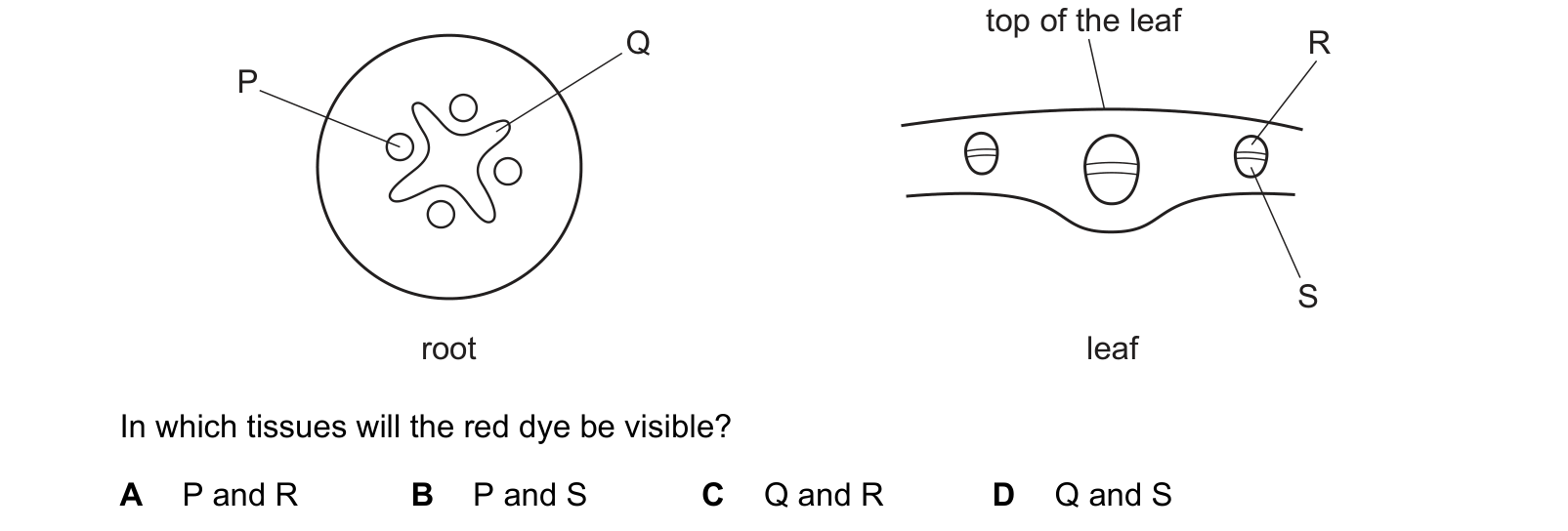
The roots of a plant were placed in a solution of red dye. After 24 hours, a section of root and a section of leaf were cut from the plant. In which tissues will the red dye be visible?
Answer: C.
In the root, xylem forms the star shape in the centre, which is Q. The small round patches between the arms of that star, one of which is P, are phloem.
In the leaf, the vein carries both tissues, with xylem on the upper side and phloem below. R points to the upper part and S to the lower, so R is the xylem.
Q and R. The positions are worth memorising because they differ by organ: in a root the xylem is a central star, in a stem it is the inner half of each bundle, and in a leaf it is the upper half of the vein. Xylem is always on the side nearest the centre or the top.
Question 2
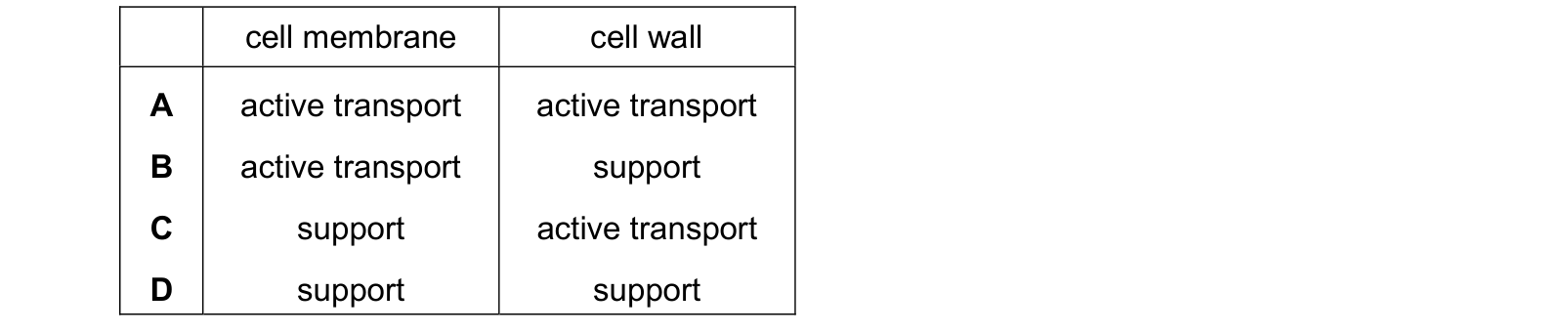
Which row shows a function of the cell membrane and a function of the cell wall in a palisade cell? Each answer gives, in order: cell membrane; cell wall.
Answer: B.
The cell membrane carries out active transport. It is partially permeable and holds the carrier proteins that move particles against a concentration gradient using energy from respiration.
The cell wall provides support. It is made of cellulose, and it is fully permeable, so it controls nothing and takes no part in transport at all. What it does is resist the outward pressure of a turgid cell, which is what holds the plant up.
Any row giving the wall a transport role has confused the two, and it is the most common error in this topic.
Question 3
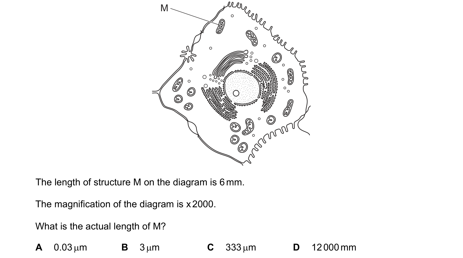
The diagram shows a human liver cell. The length of structure M on the diagram is 6 mm.
The magnification of the diagram is x 2000.
What is the actual length of M?
Answer: B.
6 ÷ 2000 = 0.003 mm
The answers are offered in micrometres, so convert: 0.003 × 1000 = 3 µm.
That figure is worth pausing on, because it is the check. A structure a few micrometres long inside a liver cell is about the size of a mitochondrion, which is exactly what it should be.
The wrong options each come from a specific slip: 0.03 µm is out by a factor of a hundred, 333 µm divides the wrong way round, and 12 000 mm multiplies instead of dividing, which would make one organelle twelve metres long.
What the syllabus asks for on this topicSyllabus points
Syllabus points
- Describe and compare the structure of a plant cell and an animal cell.
- State the functions of the structures found in cells.
- Relate the structure of specialised cells to their functions.
- Describe the levels of organisation: cell, tissue, organ, organ system, organism.
- Calculate magnification and actual size using the formula, and convert between millimetres and micrometres.
Related CIE 0610 Biology topics
Not the topic you were looking for? Describe what you are stuck on in your own words and we will take you to the notes that answer it.