Transport in animals
Contents: 7 sections
Single and double circulation
A double circulation means blood passes through the heart twice for each complete circuit of the body.
- Pulmonary circulation — heart to lungs and back.
- Systemic circulation — heart to the rest of the body and back.
Fish have a single circulation: blood passes through the heart once per circuit, going heart → gills → body → heart.
The advantage of a double circulation is the one to state precisely. Blood loses pressure passing through the narrow capillaries of the lungs. In a double circulation it returns to the heart and is re-pressurised before going to the body, so it can be delivered quickly and at high pressure. In a single circulation the blood reaching the body has already lost most of its pressure, so delivery is slower. Mammals need the faster delivery because they maintain a constant body temperature and have a high metabolic rate.
The heart
Four chambers: two atria on top, two ventricles below.
The right side handles deoxygenated blood, the left side oxygenated. Remember that on a diagram the heart is drawn as if facing you, so the right side appears on the left of the page.
The route through it:
vena cava → right atrium → right ventricle → pulmonary artery → lungs → pulmonary vein → left atrium → left ventricle → aorta → body
The pulmonary artery is the only artery carrying deoxygenated blood, and the pulmonary vein the only vein carrying oxygenated blood. That pair is asked about constantly, because it breaks the usual rule.
Why the left ventricle wall is thicker. It pumps blood to the whole body, which needs a much higher pressure, so it needs more muscle. The right ventricle pumps only to the lungs, which are close by, and too high a pressure would damage the delicate capillaries there.
Valves keep blood flowing one way. The atrioventricular valves stop backflow from ventricles into atria; the semilunar valves stop backflow from the arteries into the ventricles. A valve opens when pressure behind it is higher and closes when pressure in front is higher.
Exercise and heart rate
During activity, muscles respire faster, so they need more oxygen and glucose and produce more carbon dioxide. Heart rate and stroke volume rise, so more blood is delivered each minute, and the carbon dioxide is carried away faster.
Heart rate stays high for a while afterwards, repaying the oxygen debt built up by anaerobic respiration and clearing the lactic acid.
Coronary heart disease
The coronary arteries supply the heart muscle itself with oxygen and glucose. If fatty material builds up in their walls, the artery narrows and the blood flow falls. If the muscle is starved of oxygen it cannot respire, and a blockage causes a heart attack.
Risk factors: a diet high in saturated fat, smoking, lack of exercise, stress, obesity, age and genetic factors.
Prevention follows the same list: less saturated fat, no smoking, regular exercise, healthy weight. Treatments include stents to widen the artery and bypass surgery.
Blood vessels
| Artery | Vein | Capillary | |
|---|---|---|---|
| Direction | away from heart | towards heart | between the two |
| Wall | thick, muscle and elastic tissue | thin | one cell thick |
| Lumen | narrow | wide | very narrow |
| Valves | none | yes | none |
| Pressure | high | low | falling |
Each feature answers a need. An artery's thick elastic wall withstands and smooths the high pressure from each heartbeat. A vein's wide lumen reduces resistance where pressure is low, and valves stop backflow because there is little pressure to keep blood moving; blood in veins is helped along by the contraction of nearby skeletal muscles.
A capillary is one cell thick so the diffusion distance is short, and capillaries form dense networks so no cell is far from a supply.
Blood
Four components, each with a clear function.
- Red blood cells — carry oxygen. Packed with haemoglobin, which combines with oxygen in the lungs to form oxyhaemoglobin and releases it in respiring tissues. They are biconcave for a large surface area and have no nucleus, so they can hold more haemoglobin.
- White blood cells — defence. Phagocytes engulf and digest pathogens; lymphocytes produce antibodies.
- Platelets — fragments of cells that trigger clotting, sealing wounds to stop blood loss and to keep pathogens out.
- Plasma — the straw-coloured liquid that carries everything else: blood cells, dissolved carbon dioxide, digested food, urea, hormones and heat.
A common question shows a cell under a microscope and asks for its function. A cell with no nucleus and a pale centre is a red blood cell, carrying oxygen; a large cell with a lobed nucleus is a phagocyte, engulfing pathogens.
Check you have it
Question 1
Which row shows the features of a blood vessel that transports blood at low pressure? Each answer gives, in order: size of lumen; thickness of wall; valves.
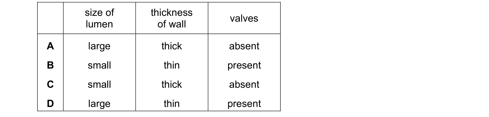
Answer: D.
All three follow from the low pressure. A thin wall is enough because there is little force to withstand. A large lumen offers little resistance to slow-moving blood. And valves are necessary precisely because low-pressure blood, often travelling against gravity, would otherwise drain backwards.
An artery is the mirror image: high pressure, so a thick wall of muscle and elastic fibres, a small lumen to maintain that pressure, and no valves, because the pressure from the ventricle keeps blood moving forwards on its own.
Each wrong row mixes the two sets. A large lumen with a thick wall, or a small lumen with valves, pairs a vein feature with an artery feature.
Blood in the veins is pushed along mainly by surrounding skeletal muscles squeezing them, and the valves turn that squeeze into one-way movement.
Question 2
What are methods for monitoring the activity of the heart? Each answer gives, in order: ECG; measuring pulse rate; listening to sounds of valves closing.
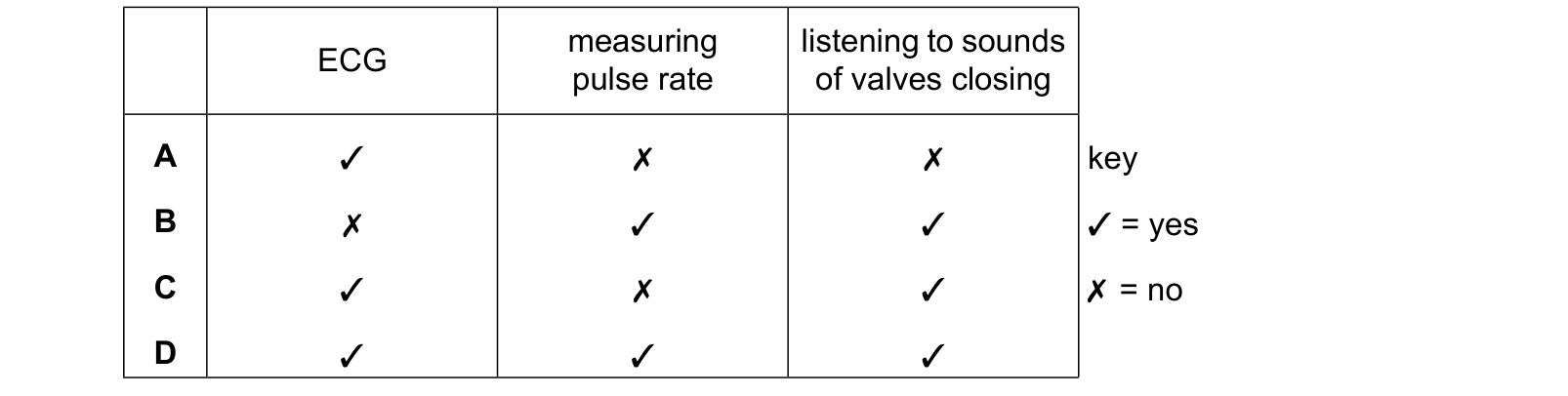
Answer: D.
An ECG, or electrocardiogram, records the electrical activity that makes the heart contract. It shows the rate and also the rhythm, so it can reveal an irregular beat that a pulse count alone would miss.
Measuring the pulse rate counts the surges of blood passing through an artery near the surface, at the wrist or neck. Each surge is one ventricular contraction, so the pulse rate is the heart rate.
Listening to the sounds of the valves closing is done with a stethoscope. The familiar two-part beat is the atrioventricular valves closing and then the semilunar valves closing, and an unusual sound, a murmur, can indicate a valve that is not sealing properly.
Each method reveals something different: electrical activity, rate, and the mechanical working of the valves. That is why all three remain in use rather than one replacing the others.
Question 3
The flow diagram shows stages in blood clotting. components of blood loss blood involved in component Y and clotting circulate forms a mesh pathogen the body entry stops
What is component X?

Answer: B.
The flow chart runs from components circulating in the blood, through the conversion of X into Y, to Y forming a mesh that stops blood loss and pathogen entry.
The substance that forms a mesh is fibrin, so Y is fibrin and X is what it was made from: fibrinogen. Fibrinogen is a soluble protein dissolved in the plasma, and fibrin is insoluble, which is exactly why the conversion is needed. Nothing can form a mesh while it is dissolved.
The -ogen ending is a useful marker throughout biology for an inactive precursor waiting to be converted.
Fibrin (A) is the product rather than the starting material, so it is one step too late.
Platelets (D) are the cell fragments that trigger the process when a vessel is damaged, releasing the chemicals that cause the conversion. They come before X rather than being X.
Plasma (C) is the liquid all of this is dissolved in.
What the syllabus asks for on this topicSyllabus points
Syllabus points
- Describe the circulatory system as a system of blood vessels with a pump and valves.
- Explain the advantages of a double circulation.
- Describe the structure and function of the heart and the effect of physical activity on heart rate.
- Describe coronary heart disease, its risk factors and its prevention.
- Describe the structure and functions of arteries, veins and capillaries.
- List the components of blood and state their functions.
Related CIE 0610 Biology topics
Not the topic you were looking for? Describe what you are stuck on in your own words and we will take you to the notes that answer it.