Five past-paper questions are below. Answer on the page: each one is marked the moment you pick, the correct option is shown whether or not you found it, and the full explanation opens either way.
CIE 0654 Co-ordinated SciencesPaper 1 and Paper 2 MCQsFree account
Organisation of the organism: five questions to try now
Real past-paper questions, the answer key from the mark scheme, and the explanation that goes with it. No account needed to answer them.
Question 1
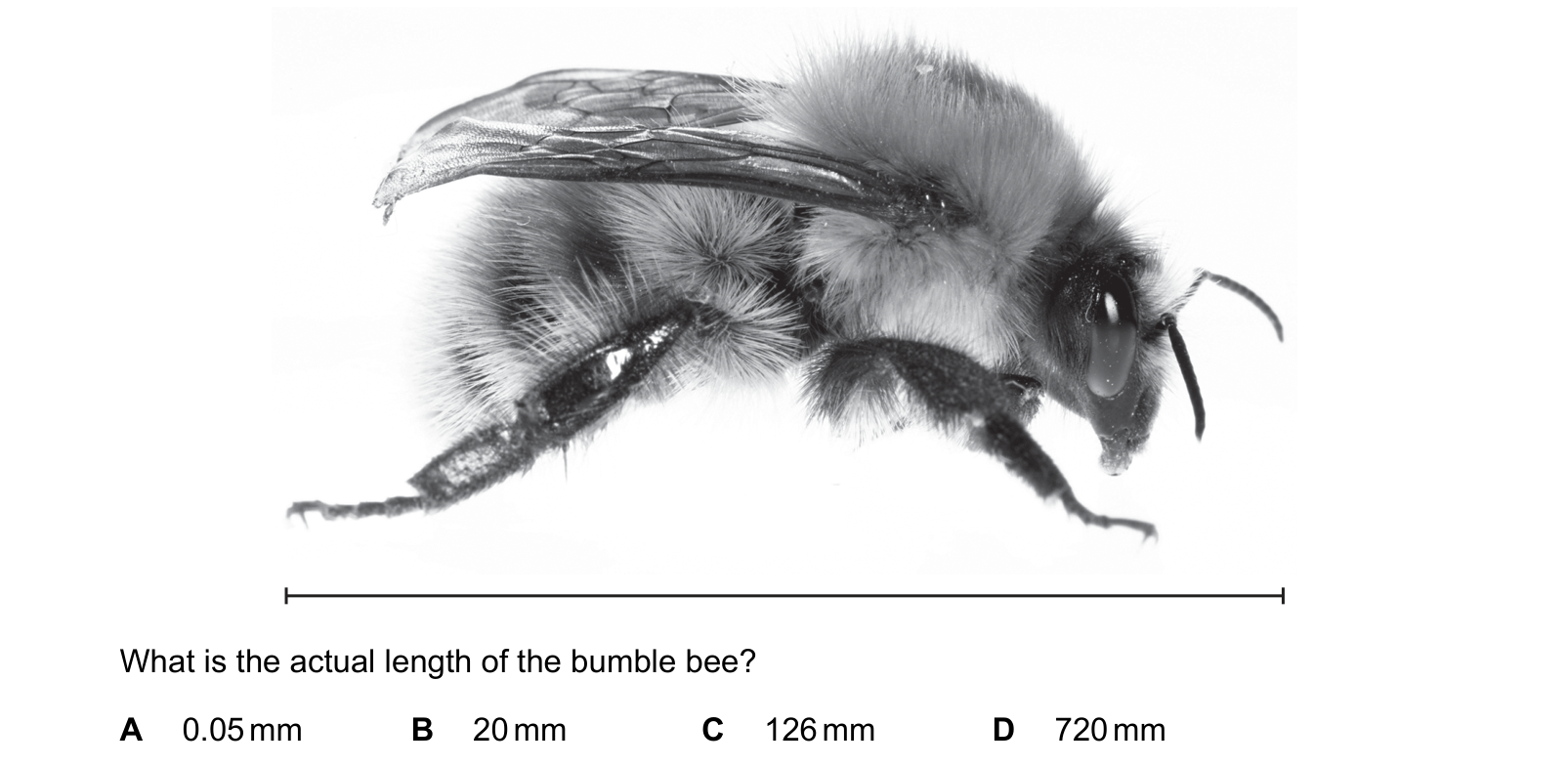
The photograph shows a bumble bee at a magnification of ×6. The line shows the length of the bumble bee. What is the actual length of the bumble bee?
Answer: B.
Magnification is the size of the image divided by the actual size, so the actual size is found by dividing the measured length by the magnification. The line drawn under the photograph is six times as long as the bee really is, and dividing it by 6 gives 20 mm, which is also a sensible length for a bumble bee. The answer of 720 mm multiplies by the magnification instead of dividing by it, which would make the insect longer than a human arm. The answer of 126 mm comes from adding the 6 to the measured length rather than dividing, and no magnification calculation ever adds. The answer of 0.05 mm is far below anything the naked eye can see and belongs to the scale of a single cell under a microscope, not to an animal photographed whole.
Question 2
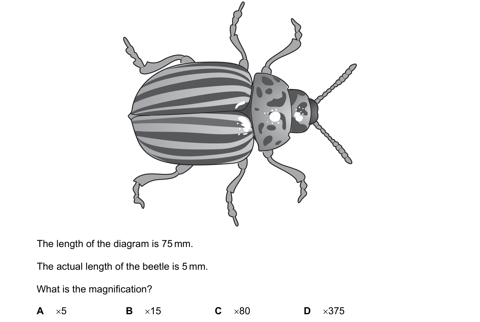
The diagram shows a beetle. The length of the diagram is 75 mm. The actual length of the beetle is 5 mm. What is the magnification?
Answer: B.
Magnification is the size of the image divided by the real size of the object, and both lengths are already in millimetres, so no conversion is needed. Dividing gives 75/5 = 15, so the drawing is fifteen times life size and B is correct. A, five, simply quotes the actual length of the beetle and calls it a magnification, which confuses the object with the ratio. D, 375, multiplies 75 by 5 instead of dividing, an error the units expose immediately, since magnification has no unit while that product would be square millimetres. C, 80, adds the two lengths together, which corresponds to no quantity at all.
Question 3
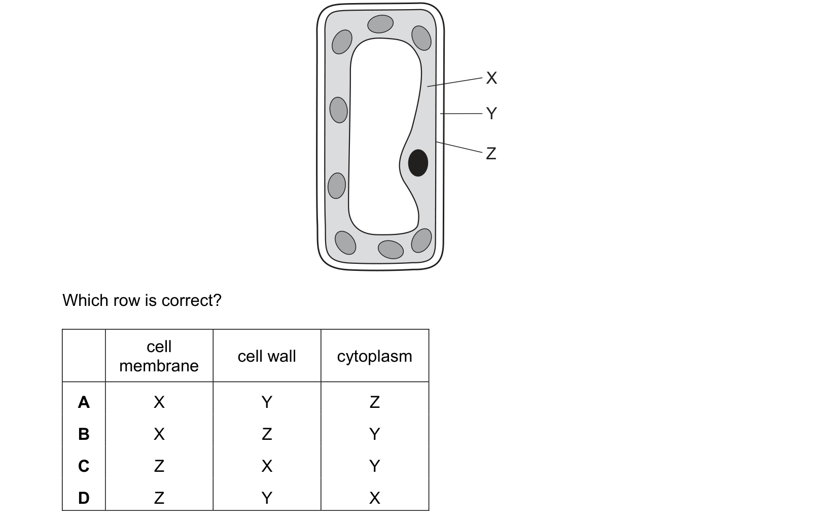
The diagram shows a typical plant cell. Which row is correct? Each answer gives, in order: cell membrane; cell wall; cytoplasm.
Answer: D.
A plant cell is built in layers from the outside inwards: the cellulose cell wall, then the cell membrane pressed against it, then the cytoplasm, and inside that the large permanent vacuole. Reading the three labels in that order gives Y on the outermost line, the cell wall, Z on the thin line just inside it, the cell membrane, and X in the granular layer that holds the chloroplasts, the cytoplasm. The two rows that call X the cell membrane place the membrane inside the cytoplasm, when the membrane is the boundary enclosing it. The row that names Z the membrane correctly but then calls X the cell wall buries the wall deep inside the cell, whereas a wall can only lie outside the membrane. Fixing the wall as the outermost layer sorts all four rows at once.
Question 4
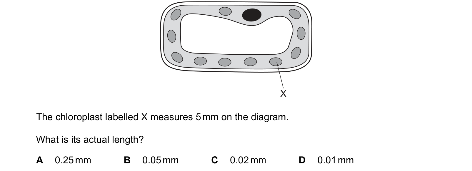
The diagram shows a palisade cell from a leaf magnified ×250. The chloroplast labelled X measures 5 mm on the diagram. What is its actual length?
Answer: C.
Actual size is the measured image size divided by the magnification, so the 5 mm chloroplast on the diagram stands for 5/250 mm of real leaf. That division gives 0.02 mm, which is 20 micrometres, a sensible length for a chloroplast. B, 0.05 mm, comes from dividing by 100 instead of 250, rounding the magnification to a convenient hundred rather than using the figure given. D, 0.01 mm, divides by 500, which doubles the stated magnification. A, 0.25 mm, is more than ten times too large, and a structure that size would be visible without any microscope.
Question 5
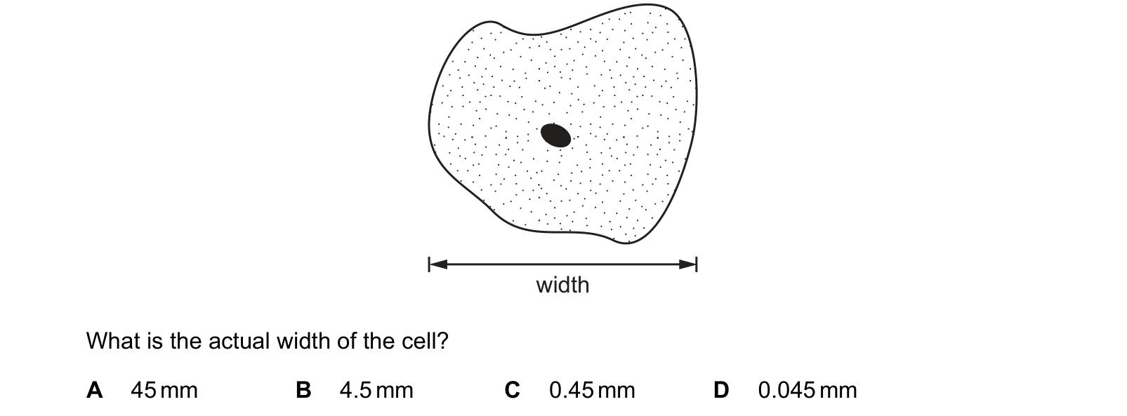
The diagram shows a cell with a magnification of ×1000. The width of the image is 45 mm. What is the actual width of the cell?
Answer: D.
Actual size is the size of the image divided by the magnification, so the width is 45 mm divided by 1000, which gives 0.045 mm, about 45 micrometres and a typical width for a cell. The answer of 45 mm leaves the measured image width untouched and would make one cell as wide as a matchbox. The answer of 4.5 mm divides by 10 and the answer of 0.45 mm divides by 100, so each has lost places while dividing by a thousand. Checking the scale of the answer settles it quickly, because cells are measured in micrometres and only 0.045 mm is small enough to need a microscope to see.
These questions are drawn from past CIE 0654 Co-ordinated Sciences papers. You answer, you find out immediately whether you were right, and you get the reasoning for the correct option and for each distractor. Wrong answers go to a mistakes locker so you can come back to exactly those.
Practice is free. You need an account only so your progress and your mistakes are still there next time.
These are the errors that cost marks on organisation of the organism, taken from our own topic notes. Read them before you practise and you will recognise the traps in the questions.
Saying plant cells have a cell wall instead of a cell membrane.
Saying the nucleus is the structure found only in plant cells. That line separates bacteria from plants and animals, not plants from animals.
Forgetting the words "large" and "permanent" in front of vacuole.
Giving root hair cells chloroplasts, or red blood cells a cell wall.
Saying the nucleus carries out photosynthesis because it controls the cell. Controlling a process is not the same as being the site of it.
Writing the magnification formula upside down as actual size divided by image size, which gives an answer below 1 for anything magnified.
Dividing a measurement in millimetres by one in micrometres without converting.
Calling a group of different tissues a tissue rather than an organ.