Gas exchange in humans
Contents: 7 sections
The route air takes
nose or mouth → larynx → trachea → bronchi → bronchioles → alveoli
Two errors show up in almost every version of this question. The larynx comes before the trachea, because the larynx is the voice box at the top of the windpipe. And bronchi come before bronchioles, because the tubes branch and get smaller as they go: one trachea splits into two bronchi, which divide again and again into ever finer bronchioles, ending at the air sacs.
The trachea and bronchi are held open by rings of cartilage, so they cannot collapse when the pressure inside the chest falls during a breath in.
Why the alveoli work
Gas exchange happens by diffusion at the alveoli, and every feature of them is there to make diffusion fast. The three requirements for fast diffusion are a large surface area, a short diffusion distance and a steep concentration gradient, and the alveoli deliver all three.
| Feature | Why it helps |
|---|---|
| Millions of tiny air sacs | An enormous total surface area for exchange |
| Wall one cell thick, and the capillary wall one cell thick too | The shortest possible diffusion distance |
| Dense network of capillaries with a good blood supply | Carries oxygen away and brings carbon dioxide, keeping the gradient steep |
| Ventilation by breathing | Constantly replaces the air, keeping the gradient steep from the other side |
| Moist lining | Gases dissolve before crossing, which they must do to diffuse |
The blood supply is the feature most often left out, and it is worth understanding rather than reciting. If blood sat still in the capillary it would soon fill with oxygen, the concentration on the two sides of the wall would equalise, and diffusion would stop. Flowing blood carries the oxygen away as fast as it arrives, so the concentration in the capillary stays low and the gradient never runs down. That is why a thin wall paired with a poor blood supply is a wrong answer even though half of it is right.
Oxygen diffuses from the alveolus into the blood. Carbon dioxide diffuses from the blood into the alveolus. Each gas moves down its own gradient, independently of the other.
Inspired and expired air
| Inspired air | Expired air | Why | |
|---|---|---|---|
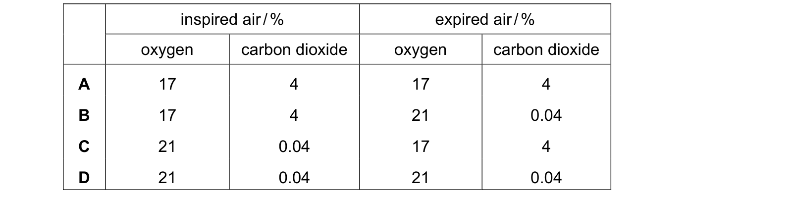
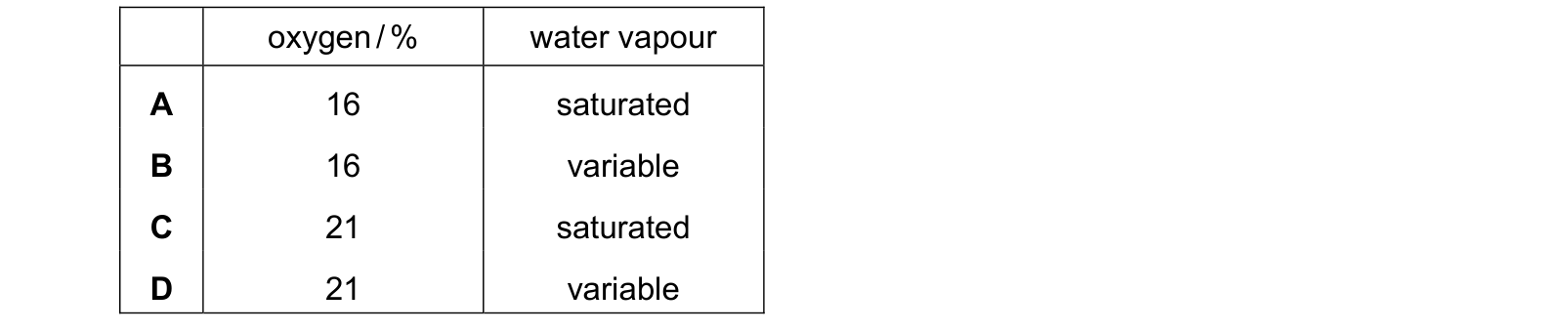
| Oxygen | About 21% | About 16% | Some has diffused into the blood |
| Carbon dioxide | About 0.04% | About 4% | Made by respiration and carried to the lungs |
| Water vapour | Variable | Saturated | The airways are warm and moist, so air picks up water on the way |
| Nitrogen | About 78% | About 78% | Not used by the body |
| Temperature | Whatever the surroundings are | Warmed to body temperature | Heat from the body |
Three things in that table decide marks.
Expired air still contains plenty of oxygen. Only about a quarter of the oxygen breathed in is absorbed, which is why mouth-to-mouth resuscitation works at all. Answering 21% for expired air is tempting because that figure is so well known, but if the oxygen were unchanged then no gas exchange would have taken place.
Water vapour is variable going in and saturated coming out. Inspired air depends on the weather, but expired air has passed over the warm wet lining of the airways every single time. That is why your breath mists a cold window even on a dry day.
Nitrogen is unchanged. It is the largest part of both, and the body does nothing with it.
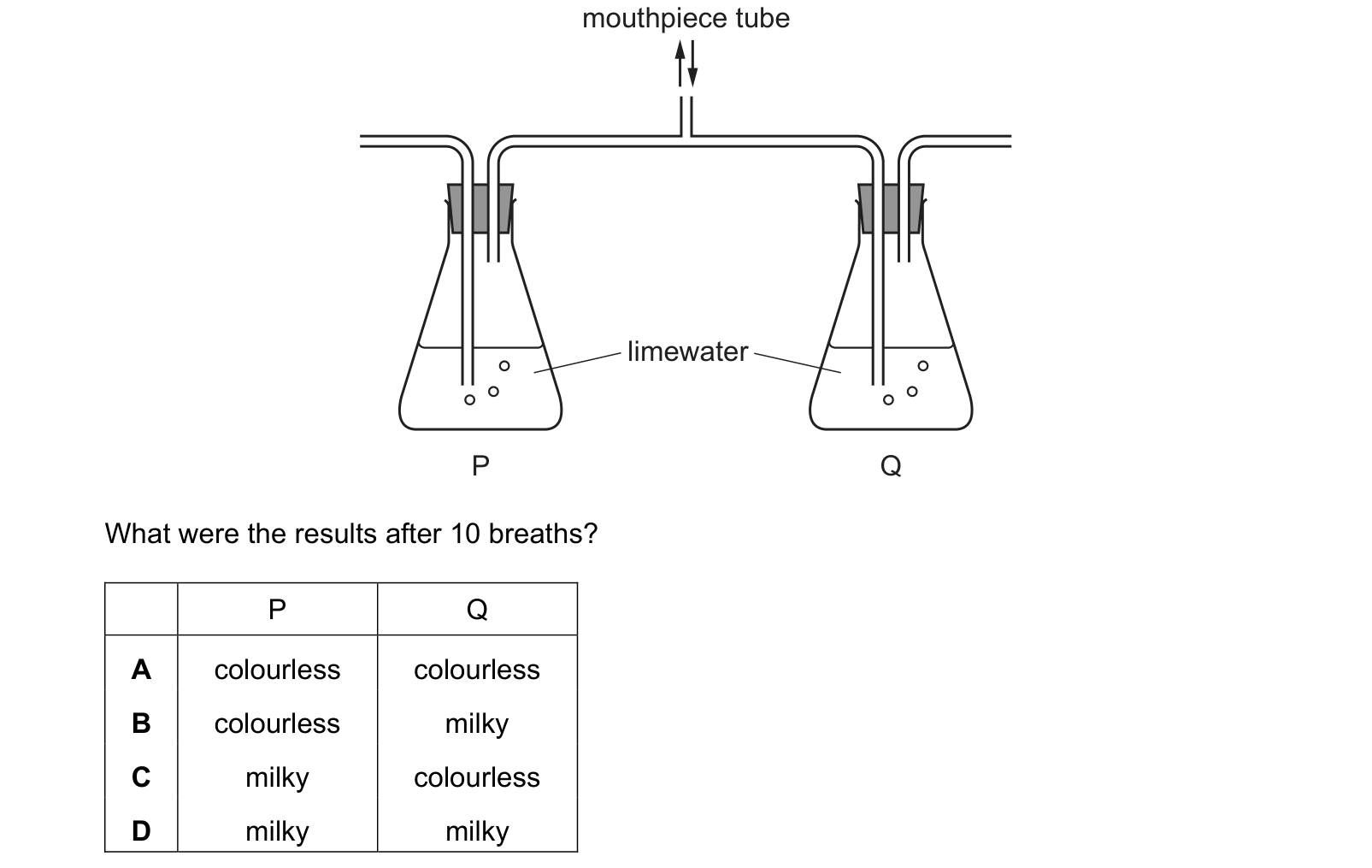
The test for the extra carbon dioxide is limewater, which turns milky. Breathing out through limewater turns it milky far faster than drawing room air through it, and that comparison is the experiment the question describes.
Cleaning the air
The airways are lined with two kinds of cell that work as a pair.
- Goblet cells make mucus. The mucus is sticky, so dust and pathogens breathed in stick to it instead of reaching the alveoli.
- Ciliated cells move the mucus. Their cilia beat in waves that sweep the loaded mucus upwards, towards the trachea and throat, where it is swallowed and destroyed by stomach acid.
Learn the pairing in exactly that order, because the question is built by swapping it: cilia do not make mucus, and mucus does not move cilia. Note the direction too. Sweeping the mucus downwards would push trapped pathogens deeper into the delicate alveoli, which is the reasoning behind that distractor.
This is also why smoking causes so much chest trouble. Smoke paralyses the cilia, so mucus is no longer cleared. It accumulates in the airways, which is the direct cause of a smoker's cough and of repeated chest infections.
Breathing and exercise
During exercise, muscles respire much faster, so they use oxygen and produce carbon dioxide much faster. The brain detects the rise in carbon dioxide in the blood and drives the diaphragm and intercostal muscles harder.
Both the rate and the depth of breathing increase. The two changes multiply together, so the volume of air moved through the lungs each minute rises far more than either change could achieve alone.
The tempting answer raises the rate but leaves the depth the same, because a faster rate is the change you notice in yourself. It is wrong for a reason worth knowing: some of every breath fills the trachea and bronchi, where no exchange happens, so shallow rapid breaths waste a larger share of each breath on air that never reaches an alveolus. Deeper breaths get more air past that dead space.
Heart rate rises at the same time and for the same reason, so that the extra oxygen is delivered and the extra carbon dioxide removed quickly. Both stay high for a while after the exercise stops, to repay the oxygen debt built up while the muscles were respiring anaerobically.
Common mistakes
- Putting the trachea before the larynx, or bronchioles before bronchi.
- Saying expired air contains no oxygen.
- Saying inspired air is dry and expired air is moist. Inspired air is variable; expired air is saturated.
- Saying nitrogen is used up in the lungs.
- Saying cilia make mucus, or that goblet cells sweep it away.
- Saying cilia move mucus down into the lungs.
- Saying alveoli walls are thin "so the gases can get through", instead of saying the diffusion distance is short.
- Leaving the blood supply out of the list of alveolar adaptations.
- Saying only the breathing rate increases during exercise.
- Calling breathing the same thing as respiration. Breathing moves air; respiration is a chemical reaction in cells.
Check you have it
Question 1
Which row is correct for inspired air and expired air? inspired air / % expired air / % oxygen carbon dioxide oxygen carbon dioxide Use the source image for W20 Paper 22, question 8.
Answer: C.
Question 2
A student breathed gently in and out of the mouthpiece of the apparatus shown. What were the results after 10 breaths? Each answer gives, in order: P; Q.
Answer: B.
Question 3
What is the expected concentration of oxygen and the water vapour content in expired air? Each answer gives, in order: oxygen / %; water vapour.
Answer: A.
What the syllabus asks for on this topicSyllabus points
Syllabus points
- Name the parts of the breathing system and trace the path of air to the alveoli.
- Describe the features of the alveoli that make them an efficient gas exchange surface.
- State the differences between inspired and expired air, and explain them.
- Describe the role of goblet cells, mucus and cilia in protecting the gas exchange system.
- Explain the effects of exercise on the rate and depth of breathing.
Related CIE 0654 Co-ordinated Sciences topics
Not the topic you were looking for? Describe what you are stuck on in your own words and we will take you to the notes that answer it.