Transport in animals
Contents: 9 sections
Double circulation
Blood passes through the heart twice for every complete circuit of the body.
- Pulmonary circulation: right side of the heart → lungs → left side of the heart.
- Systemic circulation: left side of the heart → the rest of the body → right side of the heart.
The advantage is worth stating in the form that earns the mark: blood is re-pressurised between the lungs and the body. Blood leaving the lungs has lost most of its pressure passing through the fine lung capillaries, so returning it to the heart lets it be pumped out again at high pressure. That keeps blood flowing quickly to the body, which means oxygen and glucose are delivered fast enough to support a high metabolic rate.
The naming rule
Arteries carry blood away from the heart. Veins carry blood towards the heart. The rule is about direction, not about oxygen.
That matters because of two exceptions. The pulmonary artery carries deoxygenated blood, and the pulmonary vein carries oxygenated blood. They are still an artery and a vein, because the artery leaves the heart and the vein arrives at it. Any answer that defines an artery as "a vessel carrying oxygenated blood" falls over on these two.
Round the heart
The right side handles deoxygenated blood and the left side handles oxygenated blood.
Deoxygenated route: vena cava → right atrium → right ventricle → pulmonary artery → lungs
Oxygenated route: pulmonary vein → left atrium → left ventricle → aorta → body
Blood always goes atrium first, then ventricle, on both sides.
Worked example. A red blood cell has just unloaded carbon dioxide at the lungs. Trace its route to the kidney.
pulmonary vein → left atrium → left ventricle → aorta → renal artery → kidney
The trap in this question is the last step. The kidney is being supplied, so the blood arrives in the renal artery. The renal vein takes blood away from the kidney, back towards the vena cava. Similarly, carbon dioxide leaving the body travels pulmonary artery → capillary → alveolus → bronchiole, in that order, because it crosses into the air space first and only then joins the airways.
The left ventricle has the thickest wall, because it pumps blood all the way round the body, against much greater resistance than the short trip to the lungs. The right ventricle pumps the same volume, just at lower pressure, so the difference in wall thickness is about pressure and not about volume. The septum between the two sides keeps oxygenated and deoxygenated blood from mixing.
Valves
There are two sets:
- Atrioventricular valves, between each atrium and its ventricle.
- Semilunar valves, between each ventricle and the artery leaving it.
Valves are opened and closed by pressure differences alone. They have no muscle of their own. Blood pushes a valve open in the direction pressure is falling, and pushes it shut when the pressure behind it becomes the greater.
| Stage | Atrioventricular valves | Semilunar valves |
|---|---|---|
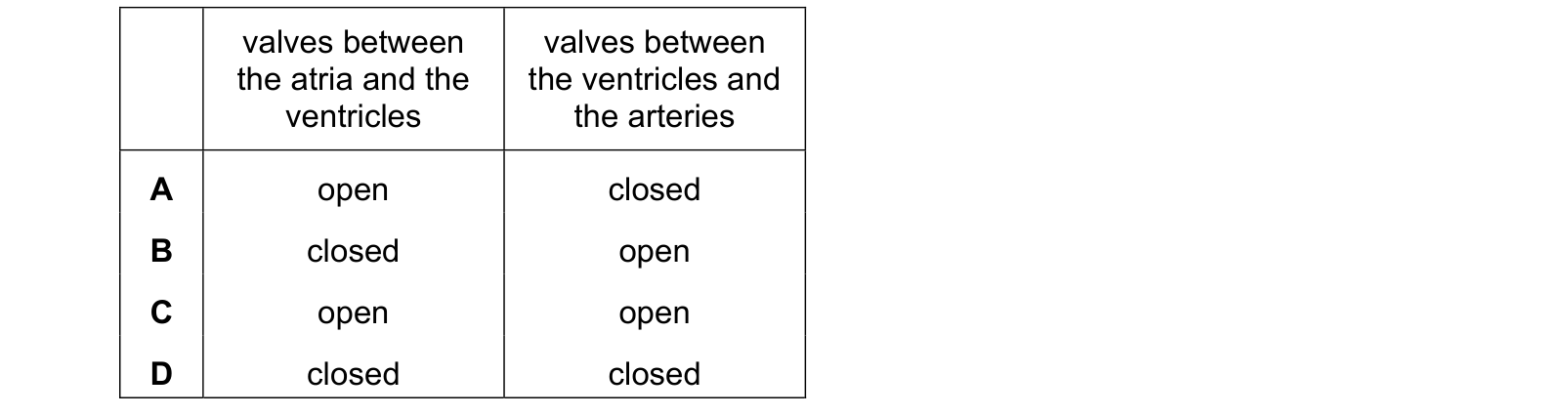
| Atria contract, ventricles relaxed and filling | Open | Closed |
| Ventricles contract | Closed | Open |
Work it out rather than memorising it. When the ventricles contract, the pressure inside them shoots above the pressure in the atria, so the atrioventricular valves slam shut, and above the pressure in the arteries, so the semilunar valves are forced open and blood is ejected. That slam is the first heart sound.
The tempting wrong answer has both sets closed, on the reasoning that this would prevent all backflow. It would also leave a powerful contraction with nowhere at all to send the blood, which is the check to apply.
Blood vessels
| Artery | Vein | Capillary | |
|---|---|---|---|
| Wall | Thick, muscular and elastic | Thin, less muscle | One cell thick |
| Lumen | Narrow | Wide | Very narrow, one red cell wide |
| Pressure | High | Low | Falling |
| Valves | None | Yes | None |
| Job | Carry blood from the heart | Return blood to the heart | Exchange with the tissues |
Every difference follows from the pressure. An artery has thick elastic walls because it has to withstand the surge each time the ventricle contracts, and the elastic recoil between beats smooths the flow. A vein carries blood at low pressure, so it needs no thick wall, but low pressure also means the blood could easily slide backwards, which is why veins have valves and arteries do not. Nearby skeletal muscles squeeze the veins and help push the blood along.
A capillary wall is one cell thick so that the diffusion distance between blood and tissue is as short as possible. Capillaries are also very numerous and narrow, which gives an enormous total surface area and slows the blood down, giving more time for exchange.
The blood
| Component | Function |
|---|---|
| Red blood cells | Carry oxygen, using haemoglobin |
| White blood cells | Defend against pathogens, by phagocytosis and by making antibodies |
| Platelets | Fragments that start blood clotting |
| Plasma | The liquid that carries the cells, plus dissolved glucose, amino acids, urea, hormones, carbon dioxide, ions and heat |
Red blood cells are packed with haemoglobin, which combines with oxygen in the lungs to form oxyhaemoglobin and releases it again in the tissues where the oxygen concentration is low. A red blood cell has no nucleus, which leaves more room for haemoglobin, and its biconcave shape gives it a larger surface area for loading and unloading oxygen.
Clotting matters for two reasons: it stops blood loss, and the scab seals the wound so pathogens cannot get in.
Matching questions on this table are usually built by swapping two functions, so the pairings to be careful with are platelets with oxygen transport, red cells with hormones, and plasma with clotting. Plasma is the closest of those to being right, because the proteins that build a clot are indeed dissolved in it, but the component whose function is clotting is the platelet.
Coronary heart disease
The coronary arteries supply the heart muscle itself with oxygen and glucose. Fatty deposits build up inside them, the lumen narrows, and blood flow to the heart muscle is reduced. If a vessel is blocked completely the muscle beyond it is starved of oxygen and dies, which is a heart attack.
Risk factors: a diet high in saturated fat, a diet high in salt, smoking, lack of exercise, stress, being overweight, age, being male, and a family history. High blood pressure is a risk factor. Low blood pressure is not, and it appears in questions precisely because "blood pressure" is remembered without the direction attached.
Reducing the risk: eat less saturated fat and salt, stop smoking, exercise regularly, keep to a healthy weight, and reduce stress. Treatments include drugs to lower blood pressure or cholesterol, stents to hold a narrowed artery open, and bypass surgery.
Common mistakes
- Defining an artery as a vessel carrying oxygenated blood, which fails for the pulmonary artery.
- Saying the pulmonary vein carries deoxygenated blood.
- Sending blood to an organ down a vein, for example writing "aorta to renal vein".
- Saying the left ventricle is thicker because it pumps more blood. It pumps at higher pressure.
- Saying valves are opened by muscles rather than by pressure differences.
- Saying both sets of valves are closed while the ventricles contract.
- Giving arteries valves.
- Saying capillary walls are thin "to let blood through". They are one cell thick to shorten the diffusion distance.
- Saying red blood cells have no nucleus so they can carry oxygen, without saying that the space is used for haemoglobin.
- Listing low blood pressure as a risk factor for coronary heart disease.
Check you have it
Question 1
The arrows in the diagram represent blood vessels. Which vessel is the pulmonary vein? Use the source image for M23 Paper 22, question 7.

Answer: B.
Question 2
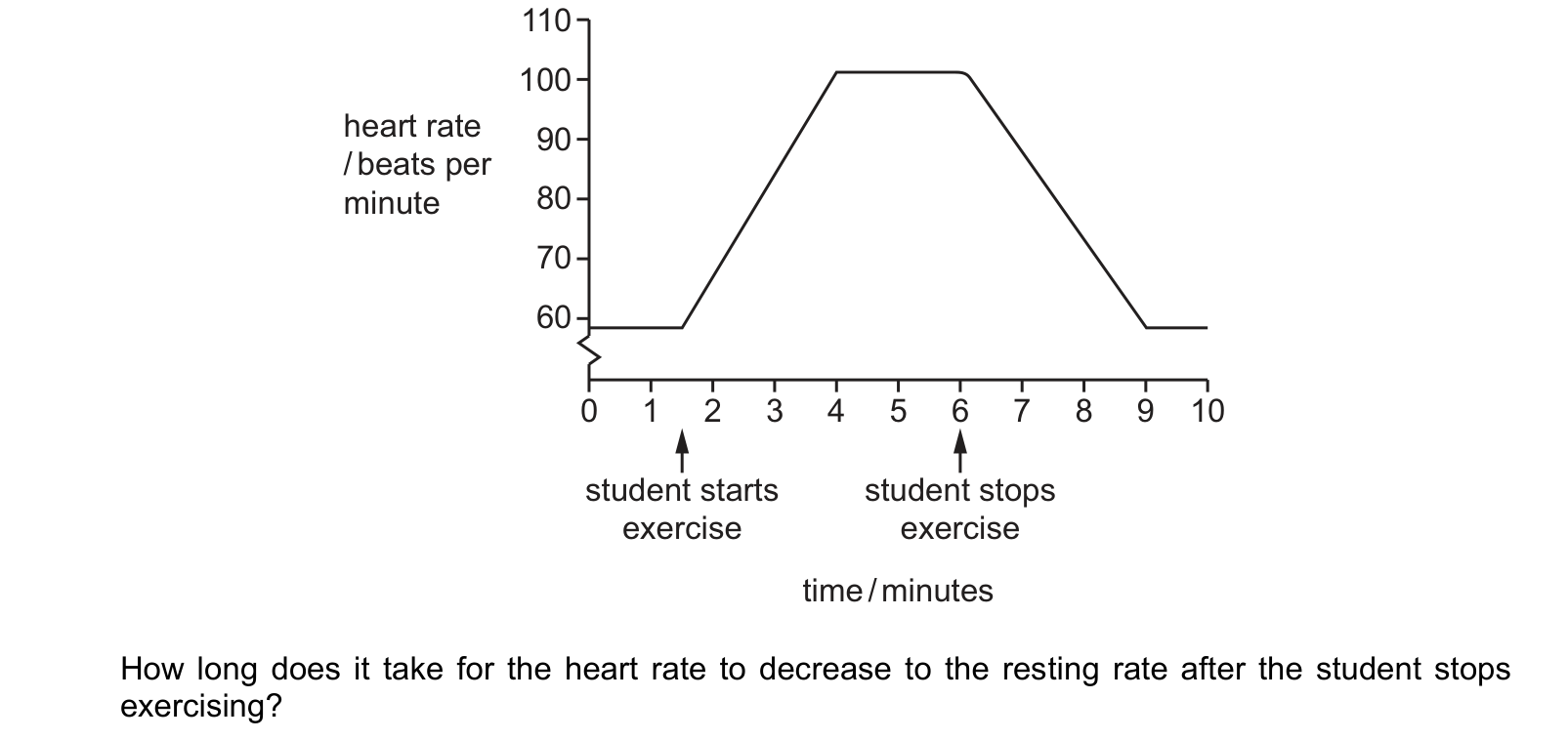
Students investigate the effect of exercise on heart rate.
The graph shows the results. exercise exercise
time / minutes
How long does it take for the heart rate to decrease to the resting rate after the student stops exercising?
Answer: A.
Question 3
Which row shows the state of the heart valves whilst the atria are contracting and the ventricles are relaxing? Each answer gives, in order: valves between the atria and the ventricles; valves between the ventricles and the arteries.
Answer: A.
What the syllabus asks for on this topicSyllabus points
Syllabus points
- Describe the double circulation of a mammal and explain its advantage.
- Name the chambers of the heart and the main blood vessels entering and leaving it.
- Describe the function of the heart valves and explain what opens and closes them.
- Compare the structure and function of arteries, veins and capillaries.
- List the components of blood and state the function of each.
- State the risk factors for coronary heart disease and how it can be reduced.
Related CIE 0654 Co-ordinated Sciences topics
Not the topic you were looking for? Describe what you are stuck on in your own words and we will take you to the notes that answer it.